

PABPC1 promotes cell proliferation and metastasis in pancreatic adenocarcinoma by regulating COL12A1 expression
- PMID: 37506150
- PMCID: PMC10336663
- DOI: 10.1002/iid3.919
PABPC1 promotes cell proliferation and metastasis in pancreatic adenocarcinoma by regulating COL12A1 expression
Abstract
Background: The expression of cytoplasmic poly (A) binding protein-1 (PABPC1) has been reported in multiple cancer types. This protein is known to modulate cancer progression. However, the effects of PABPC1 expression in pancreatic adenocarcinoma (PAAD) have not been investigated. Here, we investigate the regulatory targets and molecular mechanisms of PABPC1 in PAAD.
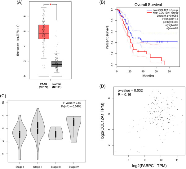
Methods: PABPC1 and collagen type XII α1 chain (COL12A1) expression in PAAD and their role in tumor prognosis and tumor stage were investigated using The Cancer Genome Atlas database analysis. After silencing PABPC1, messenger RNA sequencing and Gene Ontology (GO) and Kyoto Encyclopedia of Genes and Genomes (KEGG) analyses were performed. The expression of differentially expressed genes (DEGs), cell viability, apoptosis, and cell migration and invasion were explored using reverse transcription-quantitative polymerase chain reaction, Cell Counting Kit-8 assay, flow cytometry assay, and transwell assay, respectively. The relationship between PABPC1 and COL12A1 expression was assessed by Pearson's correlation analysis. The regulatory function of COL12A1 in PABPC1-affected BXPC3 cell behavior was studied after COL12A1 was overexpressed.
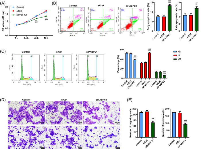
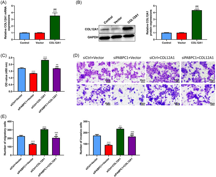
Results: PABPC1 and COL12A1 expression was upregulated in patients with PAAD and was linked to poor prognosis. Four hundred and seventy-four DEGs were observed in BXPC3 cells after PABPC1 silencing. GO and KEGG analyses revealed that the top 10 DEGs were enriched in cell adhesion pathways. Additionally, PABPC1 silencing inhibited cell viability, migration, and invasion and accelerated apoptosis in BXPC3 cells. PABPC1 silencing increased AZGP1 and ARHGAP30 expression and decreased CAV1 and COL12A1 expression in BXPC3 cells. PABPC1 positively mediated COL12A1 expression, whereas PABPC1 knockdown induced the inhibition of BXPC3 cell proliferation, migration, and invasion.
Conclusion: The results of this study indicate that PABPC1 may function as a tumor promoter in PAAD, accelerating BXPC3 cell proliferation and metastasis by regulating COL12A1 expression.
Keywords: PABPC1; cell metastasis; collagen type XII α1 chain (COL12A1); pancreatic adenocarcinoma.
© 2023 The Authors. Immunity, Inflammation and Disease published by John Wiley & Sons Ltd.
Figures


Similar articles
-
Bioinformatics analysis identified MMP14 and COL12A1 as immune-related biomarkers associated with pancreatic adenocarcinoma prognosis.Math Biosci Eng. 2021 Jun 30;18(5):5921-5942. doi: 10.3934/mbe.2021296. Math Biosci Eng. 2021. PMID: 34517516
-
PTPN2, A Key Predictor of Prognosis for Pancreatic Adenocarcinoma, Significantly Regulates Cell Cycles, Apoptosis, and Metastasis.Front Immunol. 2022 Jan 27;13:805311. doi: 10.3389/fimmu.2022.805311. eCollection 2022. Front Immunol. 2022. PMID: 35154122 Free PMC article.
-
Screening and validating the core biomarkers in patients with pancreatic ductal adenocarcinoma.Math Biosci Eng. 2019 Nov 6;17(1):910-927. doi: 10.3934/mbe.2020048. Math Biosci Eng. 2019. PMID: 31731384
-
Integrated bioinformatics analysis of expression and gene regulation network of COL12A1 in colorectal cancer.Cancer Med. 2020 Jul;9(13):4743-4755. doi: 10.1002/cam4.2899. Epub 2020 May 1. Cancer Med. 2020. PMID: 32356618 Free PMC article.
-
Identify potential prognostic indicators and tumor-infiltrating immune cells in pancreatic adenocarcinoma.Biosci Rep. 2022 Feb 25;42(2):BSR20212523. doi: 10.1042/BSR20212523. Biosci Rep. 2022. PMID: 35083488 Free PMC article. Review.
Cited by
-
Identifying and validating PLAU as a potential prognostic biomarker for PDAC.Sci Rep. 2025 Apr 11;15(1):12515. doi: 10.1038/s41598-025-97629-5. Sci Rep. 2025. PMID: 40216916 Free PMC article.
-
PABPC1 silencing inhibits pancreatic cancer cell proliferation and EMT, and induces apoptosis via PI3K/AKT pathway.Cytotechnology. 2024 Jun;76(3):351-361. doi: 10.1007/s10616-024-00626-1. Epub 2024 Apr 22. Cytotechnology. 2024. PMID: 38736728 Free PMC article.
-
High-dimensional Biomarker Identification for Scalable and Interpretable Disease Prediction via Machine Learning Models.bioRxiv [Preprint]. 2024 Oct 7:2024.10.04.616748. doi: 10.1101/2024.10.04.616748. bioRxiv. 2024. Update in: Bioinformatics. 2025 May 6;41(5):btaf266. doi: 10.1093/bioinformatics/btaf266. PMID: 39416165 Free PMC article. Updated. Preprint.
-
The Multifaced Role of Collagen in Cancer Development and Progression.Int J Mol Sci. 2024 Dec 17;25(24):13523. doi: 10.3390/ijms252413523. Int J Mol Sci. 2024. PMID: 39769286 Free PMC article. Review.
-
High-dimensional biomarker identification for interpretable disease prediction via machine learning models.Bioinformatics. 2025 May 6;41(5):btaf266. doi: 10.1093/bioinformatics/btaf266. Bioinformatics. 2025. PMID: 40286292 Free PMC article.
References
-
- Siegel RL, Miller KD, Fuchs HE, Jemal A. Cancer statistics, 2021. CA Cancer J Clin. 2021;71(1):7‐33. - PubMed
-
- De Dosso S, Siebenhüner AR, Winder T, et al. Treatment landscape of metastatic pancreatic cancer. Cancer Treat Rev. 2021;96:102180. - PubMed
-
- Mizrahi JD, Surana R, Valle JW, Shroff RT. Pancreatic cancer. Lancet. 2020;395(10242):2008‐2020. - PubMed
-
- Lim SA, Hao SB, Boyd BA, et al. Opportunity costs of surgical resection and perioperative chemotherapy for locoregional pancreatic adenocarcinoma. JCO Oncol Pract. 2022;18(4):302‐309. - PubMed
Publication types
MeSH terms
Substances
LinkOut - more resources
Full Text Sources
Medical
Molecular Biology Databases
Research Materials
Miscellaneous
