. 2023 Sep;299(9):105096.
doi: 10.1016/j.jbc.2023.105096. Epub 2023 Jul 26.

PARP14 is a writer, reader, and eraser of mono-ADP-ribosylation

Affiliations

PARP14 is a writer, reader, and eraser of mono-ADP-ribosylation

Archimede Torretta et al. J Biol Chem. 2023 Sep.

Erratum in

Abstract

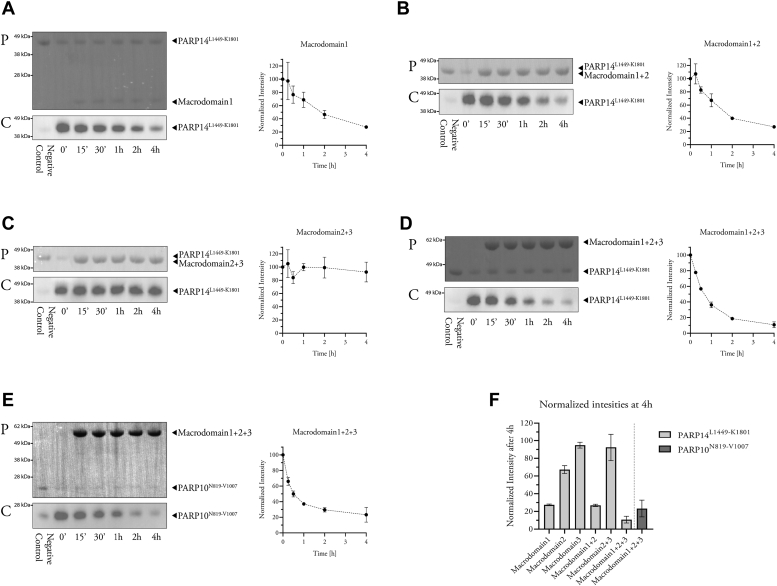
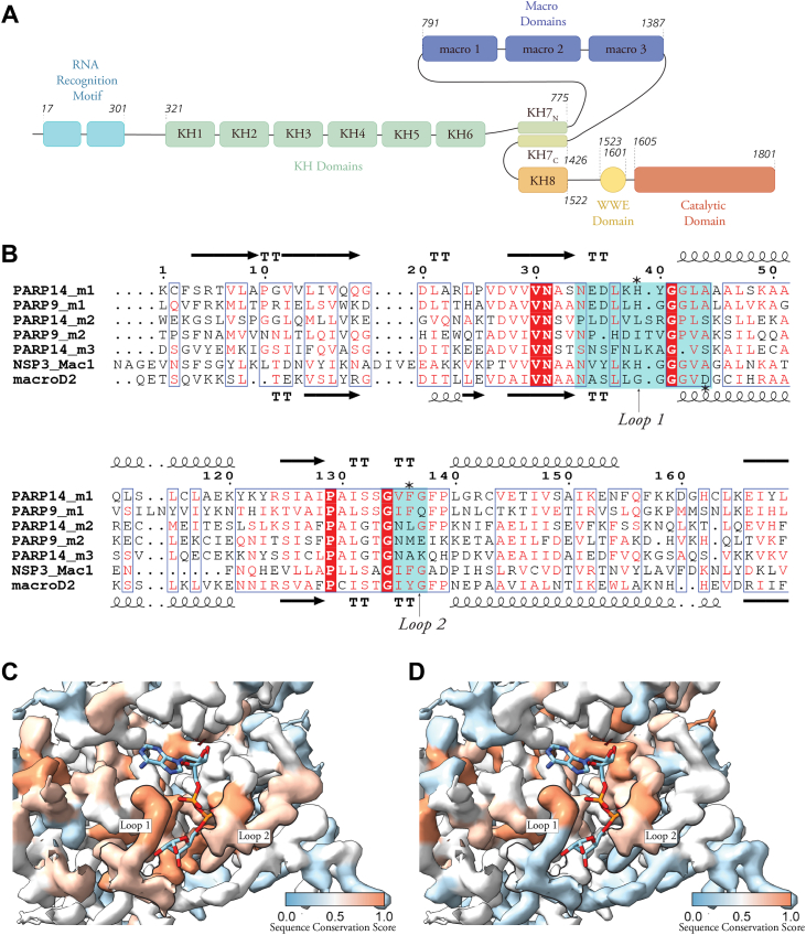
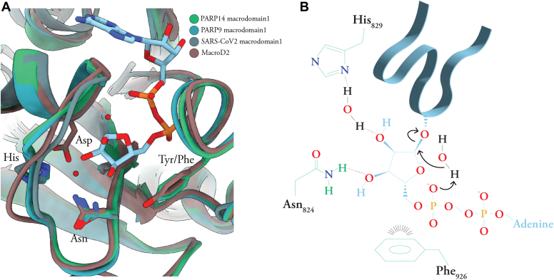
PARP14/BAL2 is a large multidomain enzyme involved in signaling pathways with relevance to cancer, inflammation, and infection. Inhibition of its mono-ADP-ribosylating PARP homology domain and its three ADP-ribosyl binding macro domains has been regarded as a potential means of therapeutic intervention. Macrodomains-2 and -3 are known to stably bind to ADP-ribosylated target proteins, but the function of macrodomain-1 has remained somewhat elusive. Here, we used biochemical assays of ADP-ribosylation levels to characterize PARP14 macrodomain-1 and the homologous macrodomain-1 of PARP9. Our results show that both macrodomains display an ADP-ribosyl glycohydrolase activity that is not directed toward specific protein side chains. PARP14 macrodomain-1 is unable to degrade poly(ADP-ribose), the enzymatic product of PARP1. The F926A mutation of PARP14 and the F244A mutation of PARP9 strongly reduced ADP-ribosyl glycohydrolase activity of the respective macrodomains, suggesting mechanistic homology to the Mac1 domain of the SARS-CoV-2 Nsp3 protein. This study adds two new enzymes to the previously known six human ADP-ribosyl glycohydrolases. Our results have key implications for how PARP14 and PARP9 will be studied and how their functions will be understood.

Keywords: ADP-ribosylation; ADP-ribosyltransferase; hydrolase; macrodomain; poly(ADP-ribose) polymerase (PARP); posttranslational modification (PTM); protein domain; signaling.


Conflict of interest statement

Conflict of interest The authors declare that they have no conflicts of interest with the contents of this article.

Figures

Figure 1
Figure 1
PARP14 macrodomain-1 displays ADP-ribosyl glycohydrolase activity in vitro.AD, automodified PARP14 catalytic domain construct was incubated with PARP14 macrodomain-1 (A), macrodomain-1+2 (B), macrodomain-2+3 (C), or macrodomain-1+2+3 (D), and the resulting levels of automodification (biotin-ADP-ribosyl) were detected using streptavidin-HRP. E, automodified PARP10 catalytic domain construct incubated with PARP14 macrodomain 1+2+3 and analyzed as above. Upper panels labeled with “P” show Ponceau S–stained membranes after transfer, while lower panels labeled with “C” show chemiluminescence images. The normalized intensities measured from each experiment are shown to the right (means ± SD of two experiments). F, comparison of the normalized intensities (means ± SD of two experiments) of the last time points of each experiment (AE). Full membranes for these experiments are shown in the Fig. S1).
Figure 2
Figure 2
PARP14 and PARP9 macrodomain-1 are related to coronavirus Nsp3 Mac1.A, schematic illustration of PARP14 domain structure with the multiple RRM and KH domains, three macrodomains, WWE domain, and the PARP-homology ADP-ribosyl transferase domain. B, sequence alignment of the indicated PARP14 and PARP9 macrodomains, macroD2, and the SARS-COV-2 Nsp3 Mac1. The secondary structures of PARP14 macrodomain-1 (Protein Data Bank: 3Q6Z) and macroD2 (4IQY) are shown above and below the alignment, respectively. Cyan boxes indicate the positions of loops 1 and 2, and asterisks mark residues referred to in the text. C, electron density from the crystal structure of PARP14 macrodomain-1 (3Q6Z) colored according to the conservation score calculated by aligning PARP14 macrodomain-1 with the catalytically active PARP9 macrodomain-1 (AlphaFold prediction), macroD2 (4IQY), and SARS-COV-2 Mac1 (6WOJ). D, electron density from the crystal structure of PARP14 macrodomain-1 colored according to the conservation score calculated by aligning PARP14 macrodomain-1 with the ADP-ribosyl binders PARP14 macrodomain-2 (3Q71), PARP14 macrodomain-3 (4ABK), and PARP9 macrodomain-2 (5AIL). Thick black outlines contour the positions of loops 1 and 2. The conservation score is a number between 0, for dissimilar residues, and 1, for identical residues.
Figure 3
Figure 3
PARP14 macrodomain-1 removes MAR from several side chains but does not break down PAR.A, percentage of ADP-ribosyl remaining on automodified PARP14 catalytic domain after treatment with macrodomains and known glycohydrolases as indicated. B, percentage of ADP-ribosyl remaining on automodified PARP10N819-V1007 after treatment with PARP14 macrodomain-1+2+3 and known glycohydrolases as indicated. C, percentage of PAR remaining on automodified PARP1 after treatment with PARP14 macrodomain-1+2+3 and glycohydrolases. D, percentage of ADP-ribosyl remaining on actin modified by C2I after treatment with PARP14 macrodomain-1 and macrodomain-1+2+3, PARP9 macrodomain 1 and glycohydrolases. All experiments, n = 4; means ± SD are shown.
Figure 4
Figure 4
Mutagenesis validates a phenylalanine residue as critical for macrodomain-1 activity.A, automodified PARP14L1449-K1801 incubated with wildtype and mutant PARP14 and PARP9 macrodomain-1. “P,” Ponceau S–stained membrane; “C,” chemiluminescence. Normalized band intensities are shown on the right. B, reactions as in A were analyzed for remaining ADP-ribosylation levels using MacroGreen. C, Differential scanning fluorimetry analysis of thermal stabilization by free ADP-ribose of both the wildtype and mutant macrodomains.
Figure 5
Figure 5
Model of macrodomain-1ADP-ribosyl glycohydrolase activity.A, superimposition of PARP14 and PARP9 macrodomain-1, macroD2, and SARS-CoV-2 macrodomain-1 with the side chains discussed in the text and the ADP-ribose molecule bound to PARP14 shown as sticks. B, schematic illustration of a possible catalytic mechanism for PARP14 macrodomain-1, based on our results as well as (38, 39, 40, 41, 42).

References

    1. Huang D., Kraus W.L. The expanding universe of PARP1-mediated molecular and therapeutic mechanisms. Mol. Cell. 2022;82:2315–2334. - PMC - PubMed
    1. Cohen M.S., Chang P. Insights into the biogenesis, function, and regulation of ADP-ribosylation. Nat. Chem. Biol. 2018;14:236–243. - PMC - PubMed
    1. Kleine H., Poreba E., Lesniewicz K., Hassa P.O., Hottiger M.O., Litchfield D.W., et al. Substrate-assisted catalysis by PARP10 limits its activity to mono-ADP-ribosylation. Mol. Cell. 2008;32:57–69. - PubMed
    1. Luscher B., Ahel I., Altmeyer M., Ashworth A., Bai P., Chang P., et al. ADP-ribosyltransferases, an update on function and nomenclature. FEBS J. 2022;289:7399–7410. - PMC - PubMed
    1. Leidecker O., Bonfiglio J.J., Colby T., Zhang Q., Atanassov I., Zaja R., et al. Serine is a new target residue for endogenous ADP-ribosylation on histones. Nat. Chem. Biol. 2016;12:998–1000. - PMC - PubMed

MeSH terms

LinkOut - more resources