. 2024 Aug;20(4):345-357.
doi: 10.1007/s11302-023-09958-7. Epub 2023 Jul 29.

The role of pyruvate-induced enhancement of oxygen metabolism in extracellular purinergic signaling in the post-cardiac arrest rat model

Affiliations

The role of pyruvate-induced enhancement of oxygen metabolism in extracellular purinergic signaling in the post-cardiac arrest rat model

Koichiro Shinozaki et al. Purinergic Signal. 2024 Aug.

Abstract

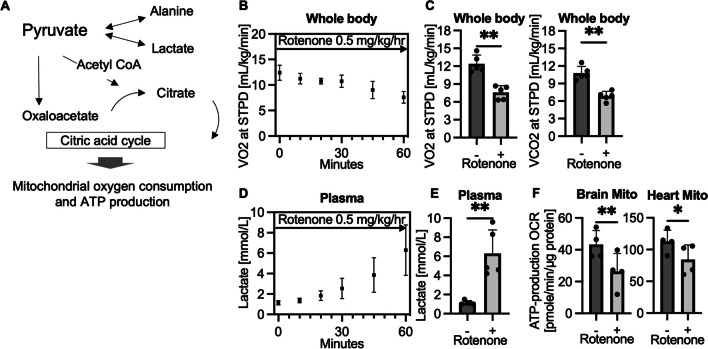
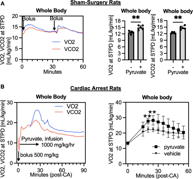
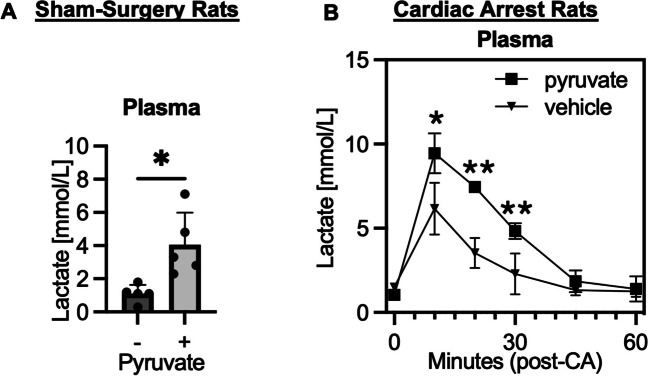
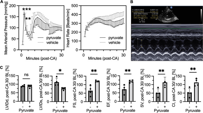
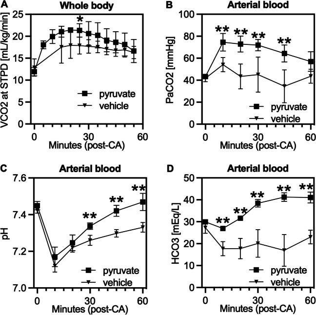
Purine nucleotide adenosine triphosphate (ATP) is a source of intracellular energy maintained by mitochondrial oxidative phosphorylation. However, when released from ischemic cells into the extracellular space, they act as death-signaling molecules (eATP). Despite there being potential benefit in using pyruvate to enhance mitochondria by inducing a highly oxidative metabolic state, its association with eATP levels is still poorly understood. Therefore, while we hypothesized that pyruvate could beneficially increase intracellular ATP with the enhancement of mitochondrial function after cardiac arrest (CA), our main focus was whether a proportion of the raised intracellular ATP would detrimentally leak out into the extracellular space. As indicated by the increased levels in systemic oxygen consumption, intravenous administrations of bolus (500 mg/kg) and continuous infusion (1000 mg/kg/h) of pyruvate successfully increased oxygen metabolism in post 10-min CA rats. Plasma ATP levels increased significantly from 67 ± 11 nM before CA to 227 ± 103 nM 2 h after the resuscitation; however, pyruvate administration did not affect post-CA ATP levels. Notably, pyruvate improved post-CA cardiac contraction and acidemia (low pH). We also found that pyruvate increased systemic CO2 production post-CA. These data support that pyruvate has therapeutic potential for improving CA outcomes by enhancing oxygen and energy metabolism in the brain and heart and attenuating intracellular hydrogen ion disorders, but does not exacerbate the death-signaling of eATP in the blood.

Keywords: Adenosine triphosphate; Energy metabolism; Gas exchange; Mitochondria; Oxygen consumption; Pyruvate.


Conflict of interest statement

Shinozaki and Becker own intellectual property of metabolic measurement in critically ill patients. Shinozaki has grant/research supported by Nihon Kohden Corp. Becker has grant/research supported by Philips Healthcare, the National Institutes of Health, Nihon Kohden Corp., BeneChill Inc., Zoll Medical Corp, Medtronic Foundation, and patents in the areas of hypothermia induction and perfusion therapies. The other authors have no disclosures.

Figures

Fig. 1
Fig. 1
Intravenous administration of mitochondrial specific inhibitor (rotenone) suppresses oxygen and energy metabolism in rats. A Schema of pyruvate metabolism (adenosine triphosphate, ATP). B Time course data of volumetric oxygen consumption (VO2) during a continuous infusion of rotenone at 0.5 mg/kg/h (sham-surgery group, n = 5 per group). C VO2 and VCO2, 60 min after a continuous infusion of rotenone as compared to base line values (average 55–60 min; sham-surgery group, n = 5 per group; **P < 0.01). D Time course data of lactate blood levels (sham-surgery group, n = 5 per group). E Blood lactate levels, 60 min after a rotenone infusion compared to base line (average 55–60 min; sham-surgery group, n = 5 per group; **P < 0.01). F ATP production in isolated mitochondria calculated from oxygen consumption rate (OCR) by Seahorse. Mitochondria were isolated from brain and heart tissues 60 min after starting the infusion of rotenone. (sham-surgery group, n = 4 per group; *P < 0.05, **P < 0.01). Values are expressed as mean +/− SD
Fig. 2
Fig. 2
Intravenous administration of mitochondrial substrate (pyruvate) increases oxygen metabolism in rats after cardiac arrest. A Representative time course data of volumetric measurements of oxygen consumption (VO2) and carbon dioxide generation (VCO2) after a bolus injections of sodium pyruvate (n = 1; STPD, standard temperature and pressure, dry). VO2 and VCO2 at 10 min after a bolus injection of pyruvate (600 mg/kg) as compared to baseline values (averaged in 5–10 min; sham-surgery group, n = 5 per group; *P < 0.05, **P < 0.01). B Post-cardiac arrest (CA) rats received a bolus injection of pyruvate (500 mg/kg) followed by a continuous infusion (1000 mg/kg/h) for 30 min. Data at time 0 was baseline before CA. Representative time course data of VO2 and VCO2 after CA (n = 1) and VO2 levels compared between the pyruvate and vehicle groups (n = 4 per group; *P < 0.05, **P < 0.01). Values are expressed as mean +/− SD
Fig. 3
Fig. 3
Intravenous administration of pyruvate increases blood lactate levels in rats after cardiac arrest. A Plasma lactate levels 10 min after a bolus injection of pyruvate (600 mg/kg) as compared to baseline values (sham-surgery group, n = 5 per group; *P < 0.05). B Post-CA rats received a bolus injection of pyruvate (500 mg/kg) followed by a continuous infusion (1000 mg/kg/h) for 30 min. Data at time 0 was baseline before CA. Plasma lactate levels compared between the pyruvate and vehicle groups (n = 4 per group; *P < 0.05, **P < 0.01). Values are expressed as mean +/− SD
Fig. 4
Fig. 4
Cardiac arrest increases blood ATP levels over time but intravenous administration of pyruvate does not increase plasma ATP levels. A Time course data of plasma ATP levels after CA (Luciferase assay, n = 8 per group; *P < 0.05, **P < 0.01). B ATP levels in plasma after pyruvate or vehicle injection compared between the non-injured (sham-surgery) and injured (post-CA) rats. (n = 6–7 per group; **P < 0.01). C ATP levels in brain and heart tissues sampled 10 min after a pyruvate (600 mg/kg) injection as compared to those in naïve rats (n = 4 per group; *P < 0.05). D ATP levels in tissues after CA collected at 30 min after a pyruvate infusion. Post-CA rats received either pyruvate or vehicle injection (n = 4 per group). Values are expressed as mean +/− SD
Fig. 5
Fig. 5
Intravenous administration of pyruvate after cardiac arrest improves cardiac contractility. Post-cardiac arrest (CA) rats received a bolus injection of pyruvate followed by a continuous infusion for 30 min. Echocardiogram was performed before CA as baseline (BL) and at 30 min after resuscitation. A Mean arterial pressure and heart rate compared between the groups (n = 8 per group; **P < 0.01, ***P < 0.001). B Representative snapshot picture of echocardiogram in our rat model. C Results of echocardiogram. LVIDd indicates diastolic left ventricular internal diameter; LVIDs, end-systolic LVID; FS, fractional shortening; EF, ejection fraction; SV, stroke volume; and CI, cardiac index. All data are calculated as percentage of values at post-CA 30 min divided by those at BL. (n = 4 per group; * P < 0.05, **P < 0.01). Values are expressed as mean +/− SD
Fig. 6
Fig. 6
Intravenous administration of sodium pyruvate induces carbon dioxide generation and attenuates acidosis after cardiac arrest. Post-CA rats received a bolus injection of pyruvate (500 mg/kg) at resuscitation followed by a continuous infusion. A VCO2, B, C, D, blood gas analysis of PaCO2, pH, and HCO3, respectively. Time 0 was at baseline before CA, and bloods were otherwise collected after resuscitation, and the values were compared between the pyruvate and vehicle injection groups (n = 4 per group; *P < 0.05, **P < 0.01). Values are expressed as mean +/− SD
Fig. 7
Fig. 7
Cardiac arrest induces purinergic signaling in the brain. A PCR assays for RNA expression of ectonucleoside triphosphate diphosphohydrolase 1 (Cd39), ecto-5′-nucleotidase (Cd73), and IL-1β (Il1b). Brain tissues were collected from rats at 2 h after CA and resuscitation and those from naïve rats. B Cd39/Cd73 expression in the heart at 72 h after CA. (n = 8–10 per group; *P < 0.05, **P < 0.01). Values are expressed as mean +/− SD

Similar articles

Cited by

References

    1. Becker LB, Aufderheide TP, Graham R (2015) Strategies to improve survival from cardiac arrest: a report from the institute of medicine. JAMA 314:223–224 10.1001/jama.2015.8454 - DOI - PubMed
    1. Merchant RM, Yang L, Becker LB, Berg RA, Nadkarni V, Nichol G, Carr BG, Mitra N, Bradley SM, Abella BS, Groeneveld PW (2011) Incidence of treated cardiac arrest in hospitalized patients in the United States. Crit Care Med 39:2401–2406 10.1097/CCM.0b013e3182257459 - DOI - PMC - PubMed
    1. Roger VL, Go AS, Lloyd-Jones DM, Adams RJ, Berry JD, Brown TM, Carnethon MR, Dai S, de Simone G, Ford ES, Fox CS, Fullerton HJ, Gillespie C, Greenlund KJ, Hailpern SM, Heit JA, Ho PM, Howard VJ, Kissela BM et al (2011) Heart disease and stroke statistics--2011 update: a report from the American Heart Association. Circulation 123:e18–e209 - PMC - PubMed
    1. Neumar RW, Eigel B, Callaway CW, Estes NA 3rd, Jollis JG, Kleinman ME, Morrison L, Peberdy MA, Rabinstein A, Rea TD, Sendelbach S (2015) The American Heart Association response to the 2015 Institute of Medicine Report on strategies to improve cardiac arrest survival. Circulation 132:1049–1070 10.1161/CIR.0000000000000233 - DOI - PubMed
    1. Nolan JP, Neumar RW, Adrie C, Aibiki M, Berg RA, Bottiger BW, Callaway C, Clark RS, Geocadin RG, Jauch EC, Kern KB, Laurent I, Longstreth WT, Merchant RM, Morley P, Morrison LJ, Nadkarni V, Peberdy MA, Rivers EP et al (2008) Post-cardiac arrest syndrome: epidemiology, pathophysiology, treatment, and prognostication: a scientific statement from the International Liaison Committee on Resuscitation; the American Heart Association Emergency Cardiovascular Care Committee; the Council on Cardiovascular Surgery and Anesthesia; the Council on Cardiopulmonary, Perioperative, and Critical Care; the Council on Clinical Cardiology; the Council on Stroke. Resuscitation 79:350–379 10.1016/j.resuscitation.2008.09.017 - DOI - PubMed

LinkOut - more resources