. 2023 Jul 28;14(1):187.
doi: 10.1186/s13287-023-03413-5.

Menstrual blood-derived mesenchymal stromal cells: impact of preconditioning on the cargo of extracellular vesicles as potential therapeutics

Affiliations

Menstrual blood-derived mesenchymal stromal cells: impact of preconditioning on the cargo of extracellular vesicles as potential therapeutics

María Ángeles de Pedro et al. Stem Cell Res Ther. .

Abstract

Background: Mesenchymal stromal cells (MSCs) have been shown to exert their therapeutic effects through the secretion of broad spectrum of paracrine factors, including extracellular vesicles (EVs). Accordingly, EVs are being pursued as a promising alternative to cell-based therapies. Menstrual blood-derived stromal cells (MenSCs) are a type of MSC that, due to their immunomodulatory and regenerative properties, have emerged as an innovative source. Additionally, new strategies of cell priming may potentially alter the concentration and cargo of released EVs, leading to modification of their biological properties. In this study, we aimed to characterize the EVs released by MenSCs and compare their therapeutic potential under three different preconditioning conditions (proinflammatory stimuli, physioxia, and acute hypoxia).

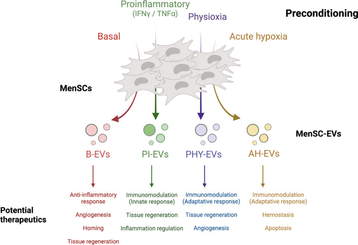
Methods: MenSCs were isolated from five healthy women. Following culturing to 80% confluence, MenSCs were exposed to different priming conditions: basal (21% O2), proinflammatory stimuli (IFNγ and TNFα, 21% O2), physioxia (1-2% O2), and acute hypoxia (< 1% O2) for 48-72 h. Conditioned media from MenSCs was collected after 48 h and EVs were isolated by a combination of ultra-filtration and differential centrifugation. An extensive characterization ranging from nano-flow cytometry (nFC) to quantitative high-throughput shotgun proteomics was performed. Bioinformatics analyses were used to derive hypotheses on their biological properties.

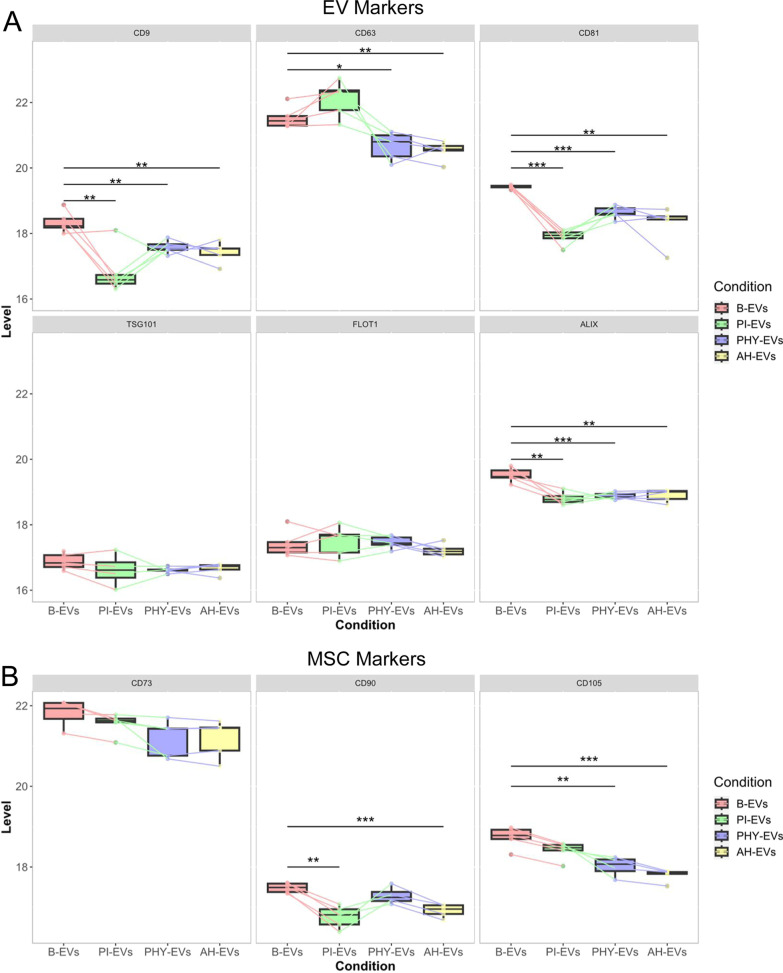
Results: No differences in the morphology, size, or number of EVs released were detected between priming conditions. The proteome analysis associated with basal MenSC-EVs prominently revealed their immunomodulatory and regenerative capabilities. Furthermore, quantitative proteomic analysis of differentially produced MenSC-EVs provided sufficient evidence for the utility of the differential preconditioning in purpose-tailoring EVs for their therapeutic application: proinflammatory priming enhanced the anti-inflammatory, regenerative and immunomodulatory capacity in the innate response of EVs, physioxia priming also improves tissue regeneration, angiogenesis and their immunomodulatory capacity targeting on the adaptive response, while acute hypoxia priming, increased hemostasis and apoptotic processes regulation in MenSC-EVs, also by stimulating immunomodulation mainly through the adaptive response.

Conclusions: Priming of MenSCs under proinflammatory and hypoxic conditions affected the cargo proteome of EVs released, resulting in different therapeutic potential, and thus warrants experimental exploration with the aim to generate better-defined MSC-derived bioproducts.

Keywords: Exosomes; Extracellular vesicles (EVs); High-throughput proteomics; Menstrual blood; Mesenchymal stromal cells (MSCs); Microvesicles; Preconditioning.


Conflict of interest statement

The authors declare that they have no competing interests.

Figures

Fig. 1
Fig. 1
Characterization of MenSC-EV samples by nFC. MenSCs were exposed to different pre-conditioning conditions for 48–72 h. Subsequently, conditioned media from MenSCs were collected in DMEM serum-free media (1%ITS, 1% P/S) for 48 h and EVs were isolated and characterized by combining ultra-filtration and differential centrifugation methods (see details in “Methods” Section). A EV release by MenSCs exposed to different priming conditions. EV release was estimated based on the number of particles released per cell exposed to basal (B, red), pro-inflammatory stimuli (PI, green), physioxia (PHY, blue) or acute hypoxia (AH, yellow) pre-conditioning. Samples from five different donor cell lines were individually analyzed by nano flow-cytometry (nFC). B Median size particles (nm) analyzed in panel A. The line indicates averaged values for n = 5 donors. C Sizing profile of representative pooled samples. A pool of n = 5 donor conditioned media samples was mixed 1:1 and EVs subsequently isolated in order to decrease inter-individual variability. Histograms represent particles detected by nFC using a bin size of 0.5 nm (small EV range, 40–200 nm). Non-linear gaussian fit curves are also plotted in the interest of visualization. EVs, extracellular vesicles; MenSCs, menstrual blood-derived stromal cells; B-EVs, EVs released by basal MenSCs; PI-EVs, EVs released by pro-inflammatory primed MenSCs; PHY-EVs, EVs released by physioxia cultured MenSCs; AH-EVs, EVs released by acute hypoxia cultured MenSCs
Fig. 2
Fig. 2
Profile of Basal MenSCs-derived EVs cargo proteome. A Characterization of the protein content of EV preparations by SDS-PAGE according to MISEV Guidelines [30]. Detection of proteins in category 1a (as tetraspanins CD63 and CD81), 2a (as TSG101, ALIX, and FLOT1), 2b (as GAPDH), and 4c (as CANX) are shown. A cell lysate, CL, (7.5 μg of total extract) was used as a control parallel to 4 × 109 particles isolated from the equally 1:1 pooled or the corresponding individual EV samples (n = 5). Molecular weights are indicated. Uncropped blots are presented in Additional file 2 and Additional file 3. GO enrichment of proteins identified in B-EVs. The most significant terms were clustered by the three GO subontologies: B Biological process (BP); C cellular component (CC); D molecular function (MF). E Reactome enrichment chart. The most significant processes are highlighted in blue and the least significant processes in red according to Benjamini-Hochberg-adjusted p values. Larger dots in the graphs indicate a greater number of proteins involved. Only the top 20 categories are shown. F Network graph obtained from Metascape composed of significantly enriched categories colored according to the functional cluster they belong to. Node size depends on the number of proteins annotated within the corresponding category. EVs, extracellular vesicles; MenSCs, menstrual blood-derived stromal cells; B-EVs, EVs released by basal MenSCs
Fig. 3
Fig. 3
Proteomic alterations in EVs were obtained following different preconditioning of MenSCs. Proteomic data of different biogroups was filtered (detection in at least three donors) and comparatively analyzed. A Principal Component Analysis (PCA) showed a high level of clustering between biogroups. B Venn diagram depicting overlapping DAPs identified in the different EV samples. DAPs identified in B-EVs (red), PI-EVs (green), PHY-EVs (blue), and AH-EVs (yellow) are represented. C Volcano plots of differentially expressed proteins in PI-EVs (left), PHY-EVs (middle), and AH-EVs (right) vs. B-EVs. Values indicate the log2FC (X-axis) and –log10adjusted p value (Y-axis). Significantly (p < 0.01) increased (red dots, log2FC ≥ 1) and decreased (green dots, log2FC ≤ -1) proteins in the preconditioning vs. basal conditions are highlighted. Top-10 dysregulated proteins are depicted on the volcano plots, together with common EV markers. DAPs: differential abundant proteins; EVs, extracellular vesicles; MenSCs, menstrual blood-derived stromal cells; B-EVs, EVs released by basal MenSCs; PI-EVs, EVs released by pro-inflammatory primed MenSCs; PHY-EVs, EVs released by physioxia cultured MenSCs; AH-EVs, EVs released by acute hypoxia cultured MenSCs
Fig. 4
Fig. 4
Changes in tetraspanins, EV biogenesis, and MSC markers in EV samples upon different preconditioning of MenSCs. A Abundance levels of the tetraspanins CD9, CD63, and CD81 (upper panel) and other EV biogenesis molecules like TSG101, FLOT1, and ALIX (lower panel) were individually evaluated among different EV groups. B The most representative MSC markers were also analyzed. Box plots indicate median protein abundance level based on MaxLFQ values in B-EVs (red), PI-EVs (green), PHY-EVs (blue), and AH-EVs (yellow) samples. Significant differences were tested by ANOVA one-way (Tukey´s post hoc test vs. basal conditions). *, p < 0.05; **, p < 0.005; and ***, p < 0.0005. EVs, extracellular vesicles; MenSCs, menstrual blood-derived stromal cells; B-EVs, EVs released by basal MenSCs; PI-EVs, EVs released by pro-inflammatory primed MenSCs; PHY-EVs, EVs released by physioxia cultured MenSCs; AH-EVs, EVs released by acute hypoxia cultured MenSCs
Fig. 5
Fig. 5
Comprehensive analysis of the MenSC-EV associated proteome according to biological function (GO ontology). A functional enrichment analysis was performed with the different DAP datasets to determine the Biological Function (BP) in which proteins were involved using DAVID. Dot-plots represent enriched GO BP terms among DAPs in PI-EVs (A), PHY-EVs (C), and AH-EVs (E) vs. B-EVs comparisons. A color-scale bar represents the level of significance for Benjamini-adjusted p values (blue, highest; red, least). The size of the dots indicates the number of proteins involved in each process. Clustering of the BP was carried out and represented in heatmaps with the most representative enriched terms from each cluster for the PI-EVs (B), PHY-EVs (D), and AH-EVs (F) vs. B-EVs comparisons. A protein score was calculated considering all proteins annotated within each category (see further details in Methods Section). The color scale of this score indicates a general up- (red) or down-regulation (green) in the preconditioning vs. basal conditions of these proteins. DAPs, differentially abundant proteins; EVs, extracellular vesicles; MenSCs, menstrual blood-derived stromal cells; B-EVs, EVs released by basal MenSCs; PI-EVs, EVs released by pro-inflammatory primed MenSCs; PHY-EVs, EVs released by physioxia cultured MenSCs; AH-EVs, EVs released by acute hypoxia cultured MenSCs
Fig. 6
Fig. 6
Reactome functional analysis. A pathway enrichment analysis was performed with the DAPs in each priming condition to determine over-represented pathways, using Reactome. A Venn diagram of the common significantly enriched pathways observed between priming conditions. Dot-plots with over-represented Reactome pathways among DAPs in PI-EVs (B), PHY-EVs (C), and AH-EVs (D) vs. B-EVs. (E) Venn diagram of DAPs among different priming conditions annotated in common immune system pathways. Voronoi diagram of the over-represented pathways within the Immune System category (R-HSA:168,256) including the DAPs from PI-EVs, green (F); PHY-EVs, purple (G); AH-EVs, yellow (H) vs. B-EVs comparisons. Dot-plots representing the top-20 significant biological processes. A color-scale bar represents the level of significance for Benjamini-adjusted p values (blue, highest; red, least). The size of the dots indicates the number of proteins involved in each process. In the voronoi diagram, a color-scale indicates the statistical significance, the lighter the more significant. Gray color is used for pathways not represented. DAPs, differentially abundant proteins; EVs, extracellular vesicles; MenSCs, menstrual blood-derived stromal cells; B-EVs, EVs released by basal MenSCs; PI-EVs, EVs released by pro-inflammatory primed MenSCs; PHY-EVs, EVs released by physioxia cultured MenSCs; AH-EVs, EVs released by acute hypoxia cultured MenSCs
Fig. 7
Fig. 7
General overview of the main findings. EVs' therapeutic potential according to the pre-selected preconditioning conditions are depicted. EVs, extracellular vesicles; MenSCs, menstrual blood-derived stromal cells; B-EVs, EVs released by basal MenSCs; PI-EVs, EVs released by pro-inflammatory primed MenSCs; PHY-EVs, EVs released by physioxia cultured MenSCs; AH-EVs, EVs released by acute hypoxia cultured MenSCs. Created with BioRender.com

Similar articles

Cited by

References

    1. Berebichez-Fridman R, Montero-Olvera PR. Sources and Clinical applications of mesenchymal stem cells: State-of-the-art review. Sultan Qaboos Univ Med J. 2018;18:e264–e277. doi: 10.18295/squmj.2018.18.03.002. - DOI - PMC - PubMed
    1. Miceli V, Bulati M, Iannolo G, Zito G, Gallo A, Conaldi PG. Therapeutic properties of mesenchymal stromal/stem cells: the need of cell priming for cell-free therapies in regenerative medicine. Int J Mol Sci. 2021;22:763. doi: 10.3390/ijms22020763. - DOI - PMC - PubMed
    1. Mutlu L, Hufnagel D, Taylor HS. The endometrium as a source of mesenchymal stem cells for regenerative medicine. Biol Reprod. 2015;92:138. doi: 10.1095/biolreprod.114.126771. - DOI - PMC - PubMed
    1. Meng X, Ichim TE, Zhong J, Rogers A, Yin Z, Jackson J, et al. Endometrial regenerative cells: a novel stem cell population. J Transl Med. 2007;5:57. doi: 10.1186/1479-5876-5-57. - DOI - PMC - PubMed
    1. Lv H, Hu Y, Cui Z, Jia H. Human menstrual blood: a renewable and sustainable source of stem cells for regenerative medicine. Stem Cell Res Ther. 2018;9:325. doi: 10.1186/s13287-018-1067-y. - DOI - PMC - PubMed

Publication types

LinkOut - more resources