Review
. 2023 Jul 3;12(7):1379.
doi: 10.3390/antiox12071379.

Oxidative Stress and Antioxidants in Age-Related Macular Degeneration

Affiliations
Review

Oxidative Stress and Antioxidants in Age-Related Macular Degeneration

Neetu Kushwah et al. Antioxidants (Basel). .

Abstract

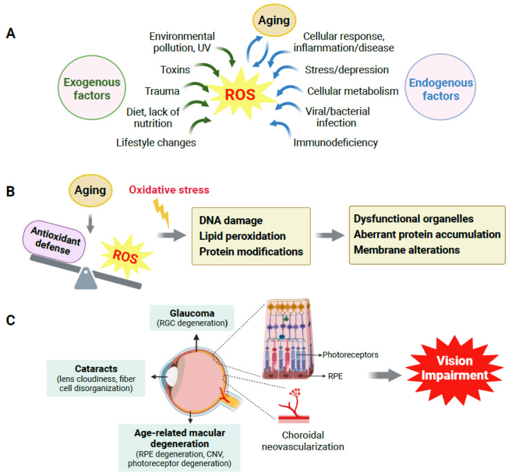
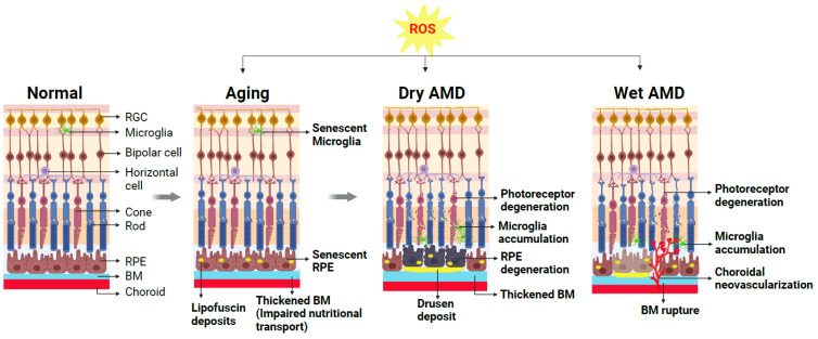
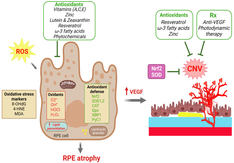
Oxidative stress plays a crucial role in aging-related eye diseases, including age-related macular degeneration (AMD), cataracts, and glaucoma. With age, antioxidant reparative capacity decreases, and excess levels of reactive oxygen species produce oxidative damage in many ocular cell types underling age-related pathologies. In AMD, loss of central vision in the elderly is caused primarily by retinal pigment epithelium (RPE) dysfunction and degeneration and/or choroidal neovascularization that trigger malfunction and loss of photo-sensing photoreceptor cells. Along with various genetic and environmental factors that contribute to AMD, aging and age-related oxidative damage have critical involvement in AMD pathogenesis. To this end, dietary intake of antioxidants is a proven way to scavenge free radicals and to prevent or slow AMD progression. This review focuses on AMD and highlights the pathogenic role of oxidative stress in AMD from both clinical and experimental studies. The beneficial roles of antioxidants and dietary micronutrients in AMD are also summarized.

Keywords: age-related macular degeneration; antioxidant; choroidal neovascularization; oxidative stress; reactive oxygen species; retinal pigment epithelium.


Conflict of interest statement

The authors declare no conflict of interest.

Figures

Figure 1
Figure 1
Aging, oxidative stress and their impact on age-related eye diseases. (A) Multiple exogenous and endogenous factors contribute to reactive oxygen species (ROS) generation, and aging plays a crucial role. (B) Oxidative stress is caused by an imbalance between ROS generation and the antioxidant defense system during aging. Oxidative stress (indicated by yellow lightning symbol) induces oxidative DNA damage, lipid peroxidation, and modifications in protein structure and functioning, which lead to dysfunctional organelles, aberrant protein accumulation, cell membrane alterations, and eventually cellular and tissue dysfunction. (C) In the eye, aging and associated oxidative stress cause oxidative damage to multiple types of ocular cells, which contribute to the onset and progression of age-related eye disorders such as age-related macular degeneration (AMD), glaucoma, and cataracts, all of which can ultimately cause vision loss in the elderly. CNV: choroidal neovascularization; RGC: retinal ganglion cell; RPE: retinal pigment epithelium. Created with BioRender.com.
Figure 2
Figure 2
Schematic representation demonstrating cellular changes in normal and aging retinas and retinas with dry AMD or wet AMD. A normal healthy retina is composed of multiple layers of retinal neurons including photoreceptors (rods and cones), bipolar cells, horizontal cells, retinal ganglion cells (RGCs), and supporting cells such as microglia, retinal pigment epithelium (RPE) cells, and choroid beneath the RPE. During normal aging, excessive reactive oxygen species (ROS) and resultant oxidative damage contribute to age-related changes in the retina (gray arrows indicate progression of retinal changes due to aging/ROS/AMD disease). Normal functions of RPE and microglia are disturbed, and these cells become dysfunctional. The accumulation of lipofuscin deposits in the RPE and impaired nutritional transport due to thickened BM are hallmarks of the aging retina, setting the stage for AMD onset. The presence of large lipid-enriched sub-RPE deposits known as drusen is a diagnostic marker for AMD. Advanced dry AMD is characterized by degenerating RPE and nearby photoreceptor degeneration. Altered choroid structure and increased microglia accumulation in the subretinal spaces indicative of chronic inflammation are also evident. In the wet form of AMD, pathological growth of abnormal choroidal blood vessels occurs through ruptured BM into the sub-RPE and subretinal space, causing exudates, hemorrhage, and thereby degeneration of the nearby photoreceptors. Created with BioRender.com.
Figure 3
Figure 3
Roles of dietary antioxidants and antioxidant defense in protecting against RPE degeneration and choroidal neovascularization (CNV) in AMD. Excessive reactive oxygen species (ROS) and oxidants cause oxidative damage in the RPE as reflected by increases in oxidative stress markers and lipid peroxidation that eventually leads to RPE atrophy. Upward pointing red arrows indicate a resultant upregulation of the indicated biomolecule. RPE dysfunction and increased VEGF production further lead to CNV formation. The effects of ROS are countered intrinsically by antioxidant reparative defense systems with antioxidant enzymes to scavenger ROS. Multiple dietary antioxidant supplements have been investigated in both clinical trials and experimental studies that have demonstrated protective effects to prevent RPE atrophy and to slow the progression of AMD. CNV formation can be treated by anti-VEGF and photodynamic therapies. Therapeutic strategies against oxidative stress by targeting Nrf2, SOD, catalase and other antioxidants may have protective effects on both RPE degeneration and CNV. Created with BioRender.com.

Similar articles

Cited by

References

    1. Phaniendra A., Jestadi D.B., Periyasamy L. Free radicals: Properties, sources, targets, and their implication in various diseases. Indian J. Clin. Biochem. 2015;30:11–26. doi: 10.1007/s12291-014-0446-0. - DOI - PMC - PubMed
    1. Hosaka S., Obuki M., Nakajima J., Suzuki M. Comparative study of antioxidants as quenchers or scavengers of reactive oxygen species based on quenching of MCLA-dependent chemiluminescence. Luminescence. 2005;20:419–427. doi: 10.1002/bio.867. - DOI - PubMed
    1. Bellezza I. Oxidative Stress in Age-Related Macular Degeneration: Nrf2 as Therapeutic Target. Front. Pharmacol. 2018;9:1280. doi: 10.3389/fphar.2018.01280. - DOI - PMC - PubMed
    1. Choo P.P., Woi P.J., Bastion M.C., Omar R., Mustapha M., Md Din N. Review of Evidence for the Usage of Antioxidants for Eye Aging. Biomed. Res. Int. 2022;2022:5810373. doi: 10.1155/2022/5810373. - DOI - PMC - PubMed
    1. Deponte M. Glutathione catalysis and the reaction mechanisms of glutathione-dependent enzymes. Biochim. Biophys Acta. 2013;1830:3217–3266. doi: 10.1016/j.bbagen.2012.09.018. - DOI - PubMed

LinkOut - more resources