Review
. 2023 Jul 7;12(7):1397.
doi: 10.3390/antiox12071397.

Antioxidant Therapy Reduces Oxidative Stress, Restores Na,K-ATPase Function and Induces Neuroprotection in Rodent Models of Seizure and Epilepsy: A Systematic Review and Meta-Analysis

Affiliations
Review

Antioxidant Therapy Reduces Oxidative Stress, Restores Na,K-ATPase Function and Induces Neuroprotection in Rodent Models of Seizure and Epilepsy: A Systematic Review and Meta-Analysis

Anderson Dutra de Melo et al. Antioxidants (Basel). .

Abstract

Epilepsy is a neurological disorder characterized by epileptic seizures resulting from neuronal hyperexcitability, which may be related to failures in Na,K-ATPase activity and oxidative stress participation. We conducted this study to investigate the impact of antioxidant therapy on oxidative stress, Na,K-ATPase activity, seizure factors, and mortality in rodent seizure/epilepsy models induced by pentylenetetrazol (PTZ), pilocarpine (PILO), and kainic acid (KA). After screening 561 records in the MEDLINE, EMBASE, Web of Science, Science Direct, and Scopus databases, 22 were included in the systematic review following the PRISMA guidelines. The meta-analysis included 14 studies and showed that in epileptic animals there was an increase in the oxidizing agents nitric oxide (NO) and malondialdehyde (MDA), with a reduction in endogenous antioxidants reduced glutathione (GSH) and superoxide dismutase (SO). The Na,K-ATPase activity was reduced in all areas evaluated. Antioxidant therapy reversed all of these parameters altered by seizure or epilepsy induction. In addition, there was a percentage decrease in the number of seizures and mortality, and a meta-analysis showed a longer seizure latency in animals using antioxidant therapy. Thus, this study suggests that the use of antioxidants promotes neuroprotective effects and mitigates the effects of epilepsy. The protocol was registered in the Prospective Register of Systematic Reviews (PROSPERO) CRD42022356960.

Keywords: Na,K-ATPase; epilepsy; exogenous antioxidants; oxidative stress.


Conflict of interest statement

The authors declare no conflict of interest.

Figures

Figure 1
Figure 1
Flow diagram of study selection. Search process using the PRISMA flow diagram.
Figure 2
Figure 2
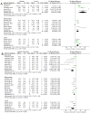
Forest plot comparing NO levels in different parts of the brain. (A) Effect of epilepsy versus control; (B) effect of intervention with exogenous antioxidants (epileptics/antioxidants versus epileptics); 95% confidence interval (CI); inverse variance (IV); standard deviation (SD); green square represents effect size. A negative standardized mean difference (SMD) represents lower levels of NO, whereas a positive SMD represents higher levels of NO [57,62,64,65,68,70].
Figure 3
Figure 3
Forest plot comparing MDA levels in different parts of the brain. (A) Effect of epilepsy versus control; (B) effect of intervention with exogenous antioxidants (epileptics/antioxidants versus epileptics); 95% confidence interval (CI); inverse variance (IV); standard deviation (SD); green square represents effect size. A negative standardized mean difference (SMD) represents lower levels of MDA, whereas a positive SMD represents higher levels of MDA [54,57,61,62,64,65,67,68,69,70,71].
Figure 4
Figure 4
Forest plot comparing GSH levels in different parts of the brain. (A) Effect of epilepsy versus control; (B) effect of intervention with exogenous antioxidants (epileptics/antioxidants versus epileptics); 95% confidence interval (CI); inverse variance (IV); standard deviation (SD); green square represents effect size. A negative standardized mean difference (SMD) represents lower levels of GSH, whereas a positive SMD represents higher levels of GSH [20,57,62,64,65,68,70].
Figure 5
Figure 5
Forest plot comparing SOD activity in different parts of the brain. (A) Effect of epilepsy versus control; (B) effect of intervention with exogenous antioxidants (epileptics/antioxidants versus epileptics); 95% confidence interval (CI); inverse variance (IV); standard deviation (SD); green square represents effect size. A negative standardized mean difference (SMD) represents lower SOD activity, whereas a positive SMD represents higher SOD activity [54,64,65,67,68,69,70,71].
Figure 6
Figure 6
Forest plot comparing CAT activity in different parts of the brain. (A) Effect of epilepsy versus control; (B) effect of Intervention with exogenous antioxidants (epileptics/antioxidants versus epileptics); 95% confidence interval (CI); inverse variance (IV); standard deviation (SD); green square represents effect size. A negative standardized mean difference (SMD) represents lower CAT activity, whereas a positive SMD represents higher CAT activity [54,57,62,67,70].
Figure 7
Figure 7
Forest plot comparing NKA activity in different parts of the brain. (A) Effect of epilepsy versus control; (B) effect of intervention with exogenous antioxidants (epileptics/antioxidants versus epileptics); 95% confidence interval (CI); inverse variance (IV); standard deviation (SD); green square represents effect size. A negative standardized mean difference (SMD) represents lower NKA activity, whereas a positive SMD represents higher NKA activity [20,54,56,57,61,62,63,64,65,67,68,69,70,71].
Figure 8
Figure 8
Effect of antioxidant use on seizure-related outcomes and mortality. (A) Seizure frequency and mortality for use of antioxidants at all dosages in the primary studies. The data are expressed as percentages. (B) Seizure latency. Forest plot comparing antioxidant-treated epileptic animals and untreated epileptic animals. Confidence interval of 95% (CI); inverse variance (IV); standard deviation (SD); green square represents effect size. A negative standardized mean difference (SMD) represents a lower latency, whereas a positive SMD represents a higher latency [20,56,62,63,64,65,68,70].

References

    1. Thijs R.D., Surges R., O’Brien T.J., Sander J.W. Epilepsy in Adults. Lancet. 2019;393:689–701. doi: 10.1016/S0140-6736(18)32596-0. - DOI - PubMed
    1. Fisher R.S., Van Emde Boas W., Blume W., Elger C., Genton P., Lee P., Engel J. Epileptic Seizures and Epilepsy: Definitions Proposed by the International League Against Epilepsy (ILAE) and the International Bureau for Epilepsy (IBE) Epilepsia. 2005;46:470–472. doi: 10.1111/j.0013-9580.2005.66104.x. - DOI - PubMed
    1. Mao X.Y., Zhou H.H., Jin W.L. Redox-Related Neuronal Death and Crosstalk as Drug Targets: Focus on Epilepsy. Front. Neurosci. 2019;13:512. doi: 10.3389/fnins.2019.00512. - DOI - PMC - PubMed
    1. Geronzi U., Lotti F., Grosso S. Oxidative Stress in Epilepsy. Expert Rev. Neurother. 2018;18:427–434. doi: 10.1080/14737175.2018.1465410. - DOI - PubMed
    1. Realmuto S., Zummo L., Cerami C., Agrò L., Dodich A., Canessa N., Zizzo A., Fierro B., Daniele O. Social Cognition Dysfunctions in Patients with Epilepsy: Evidence from Patients with Temporal Lobe and Idiopathic Generalized Epilepsies. Epilepsy Behav. 2015;47:98–103. doi: 10.1016/j.yebeh.2015.04.048. - DOI - PubMed

LinkOut - more resources