. 2023 Jul 18;12(7):1441.
doi: 10.3390/antiox12071441.

New NADPH Oxidase 2 Inhibitors Display Potent Activity against Oxidative Stress by Targeting p22phox-p47phox Interactions

Affiliations

New NADPH Oxidase 2 Inhibitors Display Potent Activity against Oxidative Stress by Targeting p22phox-p47phox Interactions

Adriana V Treuer et al. Antioxidants (Basel). .

Abstract

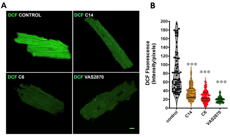
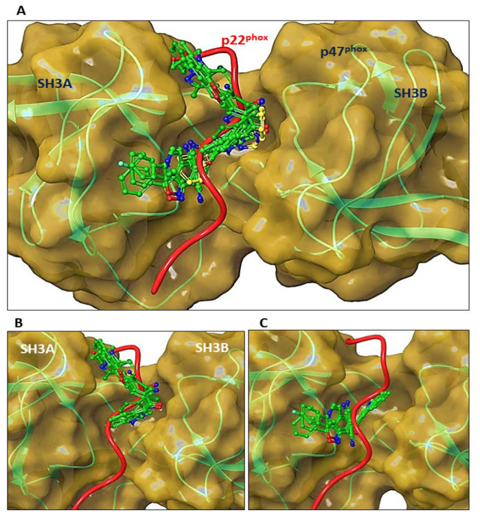
NADPH oxidase (NOX2) is responsible for reactive oxygen species (ROS) production in neutrophils and has been recognized as a key mediator in inflammatory and cardiovascular pathologies. Nevertheless, there is a lack of specific NOX2 pharmacological inhibitors. In medicinal chemistry, heterocyclic compounds are essential scaffolds for drug design, and among them, indole is a very versatile pharmacophore. We tested the hypothesis that indole heteroaryl-acrylonitrile derivatives may serve as NOX2 inhibitors by evaluating the capacity of 19 of these molecules to inhibit NOX2-derived ROS production in human neutrophils (HL-60 cells). Of these compounds, C6 and C14 exhibited concentration-dependent inhibition of NOX2 (IC50~1 µM). These molecules also reduced NOX2-derived oxidative stress in cardiomyocytes and prevented cardiac damage induced by ischemia-reperfusion. Compound C6 significantly reduced the membrane translocation of p47phox, a cytosolic subunit that is required for NOX2 activation. Molecular docking analyses of the binding modes of these molecules with p47phox indicated that C6 and C14 interact with specific residues in the inner part of the groove of p47phox, the binding cavity for p22phox. This combination of methods showed that novel indole heteroaryl acrylonitriles represent interesting lead compounds for developing specific and potent NOX2 inhibitors.

Keywords: HL-60 cells; NOX inhibitors; heteroaryl-acrylonitrile; mdx; p47phox; reactive oxygen species.


Conflict of interest statement

The authors declare no conflict of interest.

Figures

Figure 1
Figure 1
Structure of the (E)-2-(1H-Indole-3-ylcarbonyl)-3-heteroaryl-acrylonitriles studied. The reaction of cianoacetil-indole (A) with different aldehydes (B) to form the compounds studied (C1C19) is depicted. The R group for the 19 compounds (C1C19) produced is presented.
Figure 2
Figure 2
Differentiation of HL-60 cells into neutrophils, and assessment of reactive oxygen species (ROS production). (A) Western blot for gp91phox, p67phox, and p47phox NOX2 subunits. The lanes indicate homogenates of HL-60 cells untreated and HL-60 cells differentiated to neutrophils by treatment for 7 days with 1.3% DMSO. (B) Confocal microscopy image of the subunits of NOX2 present in HL-60 cells-differentiated and treated with phorbol 12-myristate 13-acetate (PMA, 0.8 µM). Green indicates the subunits gp91phox and p22phox, and red indicates the cytoplasmic subunits p47phox and p67phox. (C) Representative confocal microphotographies of ROS production in untreated and HL-60 cells differentiated to neutrophils by treatment for 7 days with 1.3% DMSO, activated with PMA 0.8 µM. (D) Graph depicting ROS production assessed by 2′,7′-dichlorofluorescein diacetate (DFC-DA) oxidation by fluorimetry. The graph indicates the change in DCF fluorescence intensity in the cells under basal conditions (not stimulated), treated with PMA, and PMA in the presence of VAS2870. Bars indicate 10 μm.
Figure 3
Figure 3
Concentration-response curves of(E)-2-(1H-Indole-3-ylcarbonyl)-3-heteroaryl acrylonitriles with NOX2 inhibition properties. The ability to inhibit NOX2-derived ROS production was assessed by monitoring in real time the fluorescence of 2′,7′-dichlorofluorescein (DCF) in HL-60 differentiated cells stimulated with phorbol 12-myristate 13-acetate (PMA, 0.8 µM) and treated with increasing concentrations of heteroaryl acrylonitriles. IC50 values are indicated for each compound. N = 4 independent experiments for each treatment.
Figure 4
Figure 4
Assessment of cytotoxicity of indole heteroaryl-acrylonitriles in neutrophil-differentiated HL-60 cells. (A); Assessment of viability (%) of HL-60 cells incubated with indole heteroaryl-acrylonitrile derivatives for 24 h. CAI; 3-cyanoacetylindole. (B), cell viability of HL-60 cells (%) after incubation for 72 h compounds C3, C6, C9, and C14. Etoposide was used to control cytotoxicity.
Figure 5
Figure 5
NOX2 inhibition evaluated in HL-60 cell membrane preparations. (A). NADPH Oxidase activity assessed by chemiluminescence. Upper panel, a graph showing NOX2 activity as relative units of luminescence per minute, measured by lucigenin chemiluminescence in membrane preparations of HL-60 cells treated with phorbol 12-myristate 13-acetate (PMA) alone (0.8 µM), PMA + VAS2870 (20 µM), and compounds C14 and C6 (both 50 µM). Lower panel: graph depicting the area under the curve (AUC) for each treatment. (B). NOX2 activity evaluated by cytochrome C reduction in membrane preparations. The bar graphs show the percentage of NOX2 activity in membrane preparations of HL-60 cells treated with increasing concentrations of compounds C14 and C6. (C). Assessment of antioxidant capacity of the indole heteroaryl-acrylonitrile compounds. The graph shows the antioxidant capacity of the new acrylonitrile derivatives, expressed as percentage of the discoloration of DPPH in a concentration range between 30–300 µM. N = 3 independent experiments for each treatment. *, p < 0.05 vs. control; ***, p < 0.0001 vs. control.
Figure 6
Figure 6
Inhibition of NOX2-derived ROS production in cardiomyocytes from mdx mice. (A). Representative confocal microphotographies of isolated mdx cardiomyocytes loaded with the ROS-sensitive probe DCF (green) fluorescent probe and treated with the compounds C6, C14 (both 1 µM) and VAS2870 (20 µM) as a control. The bar indicates 10 μm. (B). Violin graphs showing the fluorescence intensity of DCF from individual myocytes, measured as intensity per pixel. Cells for each condition were obtained from 3 mdx mice’s hearts. One-way ANOVA, ***, p < 0.0001, vs. control.
Figure 7
Figure 7
Evaluation of p47phox membrane translocation in HL-60 cells Western blot analysis of phorbol 12-myristate 13-acetate (PMA)-stimulated p47phox translocation in HL-60 cell homogenates. Left panel representative Western blot showing the translocation of p47phox from the cytosol to the membrane after treatment with PMA (0.8 µM) alone, PMA plus VAS2870 (20 µM), and PMA plus compound C6 (50 µM). Right panel: graph showing the membrane/cytosol ratio of p47phox after the pharmacological treatments. *** p < 0.0002, n = 3 independent experiments.
Figure 8
Figure 8
Molecular docking of indole heteroaryl-acrylonitriles in the groove of activated p47phox. (A): Comparison of the binding mode of C2, C3, C4, C5, C6, C9, C10, and C14 ligands (green) with the p22phox tail (red) and VAS2870 (yellow) in the binding interface of activated p47phox. (B): binding mode of the less active ligands (C2, C4, C9, and C10). (C): binding mode of the most active ligands C3, C5, C6, and C14.
Figure 9
Figure 9
(A) Overview of compound C6 (CPK representation) within the p47phox binding pocket (molecular surface) extracted from docking simulations (C6 carbon atoms in green). (B) 3D scheme of p47phox-C6 interaction in a surface representation (colored by partial atomic charge). Hydrogen bridge interactions are represented by dashed yellow lines and π-π staking by dashed light blue lines. (C) 2D interactions diagram of the docking-generated pose for compound C6, highlighting the key main interactions with amino acid residues of p47phox. Green lines depict π-π staking, magenta lines indicate hydrogen bonds.
Figure 10
Figure 10
Impact of indole heteroaryl-acrylonitriles on cardiac damage induced by ischemia-reperfusion. (A) Graph depicting the values of ventricular developed pressure in control conditions and in the presence of compounds C6 and C14, both 1 µM; and apocynin (100 µM). (B) Evaluation of compounds C6, C14, and apocynin on infarct size in the protocol of cardiac ischemia-reperfusion. The numbers of hearts used: control n = 6; C6 n = 5; C14 n = 3; apocynin n = 5. * p < 0.05, ** p < 0.01 vs. control, one- or two-way ANOVA.

Similar articles

Cited by

References

    1. Brandes R.P., Weissmann N., Schroder K. Nox family NADPH oxidases: Molecular mechanisms of activation. Free Radic. Biol. Med. 2014;76:208–226. doi: 10.1016/j.freeradbiomed.2014.07.046. - DOI - PubMed
    1. Noreng S., Ota N., Sun Y., Ho H., Johnson M., Arthur C.P., Schneider K., Lehoux I., Davies C.W., Mortara K., et al. Structure of the core human NADPH oxidase NOX2. Nat. Commun. 2022;13:6079. doi: 10.1038/s41467-022-33711-0. - DOI - PMC - PubMed
    1. Kawahara T., Quinn M.T., Lambeth J.D. Molecular evolution of the reactive oxygen-generating NADPH oxidase (Nox/Duox) family of enzymes. BMC Evol. Biol. 2007;7:109. doi: 10.1186/1471-2148-7-109. - DOI - PMC - PubMed
    1. Vermot A., Petit-Hartlein I., Smith S.M.E., Fieschi F. NADPH Oxidases (NOX): An Overview from Discovery, Molecular Mechanisms to Physiology and Pathology. Antioxidants. 2021;10:890. doi: 10.3390/antiox10060890. - DOI - PMC - PubMed
    1. Liu R., Song K., Wu J.X., Geng X.P., Zheng L., Gao X., Peng H., Chen L. Structure of human phagocyte NADPH oxidase in the resting state. Elife. 2022;11:e83743. doi: 10.7554/eLife.83743. - DOI - PMC - PubMed

LinkOut - more resources