. 2023 Jun 25;12(7):906.
doi: 10.3390/biology12070906.

Combination Effects of Integrin-linked Kinase and Abelson Kinase Inhibition on Aberrant Mitosis and Cell Death in Glioblastoma Cells

Affiliations

Combination Effects of Integrin-linked Kinase and Abelson Kinase Inhibition on Aberrant Mitosis and Cell Death in Glioblastoma Cells

Abigail Cunningham et al. Biology (Basel). .

Abstract

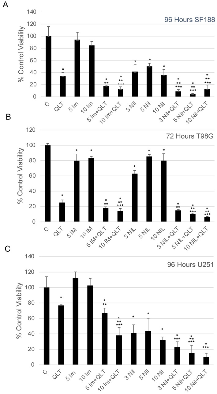
In cancer cells, inhibition of integrin-linked kinase (ILK) increases centrosome declustering causing mitotic arrest and cell death. Yet, not all cancer cells are susceptible to anti-ILK treatment alone. We investigate a combination drug strategy targeting ILK and another oncogenic kinase, Abelson kinase (ABL). Drug-concentration viability assays (i.e., MTT assays) indicate that ILK and ABL inhibitors in combination decreased the viability of glioblastoma cells over the ILK drug QLT-0267 alone. Combination strategies also increased aberrant mitoses and cell death over QLT-0267 alone. This was evident from an increase in mitotic arrest, apoptosis and a sub-G1 peak following FAC analysis. In vitro, ILK and ABL localized to the centrosome and the putative ILK kinase domain was important for this localization. Increased levels of cytosolic ABL are associated with its transformative abilities. ILK inhibitor effects on survival correlated with its ability to decrease cytosolic ABL levels and inhibit ABL's localization to mitotic centrosomes in glioblastoma cells. ILK inhibitor effects on ABL's centrosomal localization were reversed by the proteasomal inhibitor MG132 (a drug that inhibits ABL degradation). These results indicate that ILK regulates ABL at mitotic centrosomes and that combination treatments targeting ILK and ABL are more effective then QLT-0267 alone at decreasing the survival of dividing glioblastoma cells.

Keywords: Abelson kinase; apoptosis; centrosome declustering; glioblastoma; integrin-linked kinase; mitotic arrest.


Conflict of interest statement

The authors declare no conflict of interest.

Figures

Figure 1
Figure 1
ILK and ABL inhibitors in combination significantly decrease viability over individual inhibitors alone. (A) An MTT viability assay was performed after SF188 cells were exposed to QLT-0267 (10 μM) with or without imatinib (5 and 10 μM) or nilotinib (3, 5 and 10 μM) for 96 h. n = 5 separate platings, with each treatment group run in triplicate or quadruplicate and averaged. * different from vehicle C, ** different from ABL inhibitor at the same concentration and *** different from QLT alone; determined using an ANOVA and Fisher’s (LSD) test. p < 0.05. (B) An MTT viability assay was performed after T98G cells were exposed to QLT-0267 (10 μM) with or without either imatinib (5 and 10 μM) or nilotinib (3, 5 and 10 μM) for 72 h. n = 3–4 separate platings. * different from vehicle C, ** different from ABL inhibitor at the same concentration; determined using an ANOVA and Fisher’s (LSD) test. *** different from QLT alone; determined using a paired t-test. p < 0.05. (C) An MTT viability assay was performed after U251 cells were exposed to QLT-0267 (10 μM) with or without either imatinib (5 and 10 μM) or nilotinib (3, 5 and 10 μM) for 96 h. n = 3 separate platings. * different from vehicle C, ** different from ABL inhibitor at the same concentration and *** different from QLT alone; determined using an ANOVA and Fisher’s (LSD) test. p < 0.05.
Figure 2
Figure 2
ILK and ABL inhibitors in combination significantly increase G1/S cell cycle arrest and apoptosis over individual inhibitors alone. (A) FACS analysis was performed in T98G cells treated with QLT-0267 (10 μM) with or without imatinib (5 and 10 μM) for 72 h. Nonadherent and adherent cells were pooled. The percentage of cells in sub-G1, G0/G1-, S- and G2/M is expressed and an average ± SEM. * different from vehicle C; ** different from QLT alone. n = 3 separate platings. ANOVA and Fisher’s (LSD) test. p < 0.05. (B) Apoptotic cells were quantitated in T98G cells treated with QLT-0267 (10 μM) with or without imatinib (5 and 10 μM) or nilotinib (3, 5 and 10 μM) for 24 h. (C) Likewise, the average cells in the field of view (represented as a percentage of control) are shown. * different from C, QLT alone or nilotinib alone at the same concentration. n = 3–4. ANOVA and Fisher’s (LSD) test. p < 0.05.
Figure 3
Figure 3
ILK and ABL inhibitors in combination exert a synergistic effect on aberrant mitoses and mitotic arrest over individual inhibitors alone. Mitotic cells were quantitated in T98G cells after exposure to the following drugs: 10 μM QLT-0267 (QLT) and 5 or 10 μM imatinib alone (5 Im or 10 Im) or in combination and compared to drug vehicle control (Control). Cells were exposed to drug(s) or drug vehicle in complete media containing serum for 24 h, fixed and immunocytochemically stained with an α-tubulin and pericentrin antibody. Nuclei were counterstained with DAPI. (A) The percentage of mitotic cells was determined for each treatment group; 5–10 fields of view were randomly chosen and analyzed per trial. The total cell number analyzed is over 1800. QLT-0267 significantly increased the percentage of mitotic cells above vehicle control while imatinib was not different from control. QLT-0267 together with imatinib was significantly greater than QLT alone. Values were averaged and expressed as percent control ± SEM. p < 0.05, * different from control; ** different from QLT alone, n = 4. Significance was determined by an ANOVA and Fisher’s (LSD) test. (B) Representative T98G cells following exposure to 10 μM QLT-0267 (QLT) and 5 or 10 μM imatinib alone (5 Im or 10 Im) or in combination with QLT-0267. Multipolar spindles, aberrant mitotic spindles and pericentrin fragmentation were observed in the presence of QLT alone or together with imatinib. Scale bar = 10 µm. (C) Representative T98G cells following exposure to 10 μM QLT-0267 (QLT) alone or together with 5 or 10 μM nilotinib in combination. Aberrant mitotic events (i.e., multipolar spindles and pericentrin fragmentation) occur in the presence of QLT alone or together with nilotinib. Additionally, a pronounced increase in apoptotic cells (labelled with white arrows) was seen in combination treatment groups. n = 3–4. Scale bar = 10 µm.
Figure 4
Figure 4
ILK and ABL localize to the centrosome in glioblastoma cells. (A) In T98G cells, FLAG-tagged ILK-WT colocalized with pericentrin immunostaining while the ILK mutant, FLAG-tagged ILK-KD did not. (B) FLAG-tagged ABL was seen to colocalize with pericentrin while the empty control plasmid did not. Shown are representative figures of three separate platings. Large multinucleated cells undergoing cell division were frequently seen (as shown in the lower panel of the FLAG-tagged ABL tranfected cells).
Figure 5
Figure 5
The ILK inhibitor QLT-0267 decreases ABL in the cytosolic fraction and the proteasome inhibitor MG132 rescues these effects in T98G cells. (A) T98G glioblastoma cells were exposed to either DMSO or 10 μM QLT-0267 (QLT) alone or together with 1 μM or 10 μM MG132 for 6 h. Western blot analysis following cell fractionation indicates that increasing amounts of MG132 in the presence of QLT-0267 rescued cytoplasmic ABL levels. The purity of cellular fractions was verified using the primarily cytosolic (α-tubulin) and nuclear (Lamin B1) proteins. Please check the original images in Figure S1. (B) Following Western blotting, densitometric analysis of proteins was performed. Values were averaged and expressed as percent control ± SEM. p < 0.05, * different from control or QLT-0267 together with 10 μM MG132 as determined by a paired Student’s t-test, n = 5 independent trials.
Figure 6
Figure 6
The ILK inhibitor QLT-0267 decreases ABL at the centrosome in glioblastoma cells. T98G cells were exposed to drug vehicle, 10 μM QLT-0267 alone or together with increasing concentrations of MG132 (1 μM or 10 μM) for 6 h in serum-containing media. Cells were then immunocytochemically labelled using an ABL and pericentrin antibody and nuclei were counterstained with DAPI. (A) Loss of ABL immunofluorescence at the centrosome of mitotically dividing cells was observed in T98G cells exposed to 10 μM QLT-0267 and this loss was rescued by 1 μM and 10 μM of the proteosomal inhibitor MG132. White arrows highlight mitotically dividing cells. An overlay of mitotically dividing cells is shown at low (see “Overlay” above) and high magnification (see “Mag” above). Calibration bars represent 10 μm. (B) ABL immunofluorescence was quantified at mitotic centrosomes in glioblastoma cells. The calculated total cell fluorescence at the centrosome (CTCF) was determined for C, 10 μM QLT-0267 alone or together with 1 μM MG132. Data represent n = 5 (separate culture platings) for T98G cells. p < 0.05, * different from control.

References

    1. McDonald P.C., Fielding A.B., Dedhar S. Integrin-linked kinase—Essential roles in physiology and cancer biology. J. Cell Sci. 2008;121:3121–3132. doi: 10.1242/jcs.017996. - DOI - PubMed
    1. Wang J., Pendergast A.M. The Emerging Role of ABL Kinases in Solid Tumors. Trends Cancer. 2015;1:110–123. doi: 10.1016/j.trecan.2015.07.004. - DOI - PMC - PubMed
    1. Greuber E.K., Smith-Pearson P., Wang J., Pendergast A.M. Role of ABL family kinases in cancer: From leukaemia to solid tumours. Nat. Rev. Cancer. 2013;13:559–571. doi: 10.1038/nrc3563. - DOI - PMC - PubMed
    1. Zheng C.C., Hu H.F., Hong P., Zhang Q.H., Xu W.W., He Q.Y., Li B. Significance of integrin-linked kinase (ILK) in tumorigenesis and its potential implication as a biomarker and therapeutic target for human cancer. Am. J. Cancer Res. 2019;9:186–197. - PMC - PubMed
    1. Edwards L.A., Woo J., Huxham L.A., Verreault M., Dragowska W.H., Chiu G., Rajput A., Kyle A.H., Kalra J., Yapp D., et al. Suppression of VEGF secretion and changes in glioblastoma multiforme microenvironment by inhibition of integrin-linked kinase (ILK) Mol. Cancer Ther. 2008;7:59–70. doi: 10.1158/1535-7163.MCT-07-0329. - DOI - PubMed

LinkOut - more resources