. 2023 Jul 8;12(7):975.
doi: 10.3390/biology12070975.

Revision of the Western Indian Ocean Angel Sharks, Genus Squatina (Squatiniformes, Squatinidae), with Description of a New Species and Redescription of the African Angel Shark Squatina africana Regan, 1908

Affiliations

Revision of the Western Indian Ocean Angel Sharks, Genus Squatina (Squatiniformes, Squatinidae), with Description of a New Species and Redescription of the African Angel Shark Squatina africana Regan, 1908

Simon Weigmann et al. Biology (Basel). .

Abstract

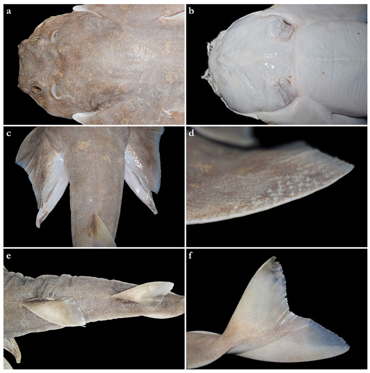
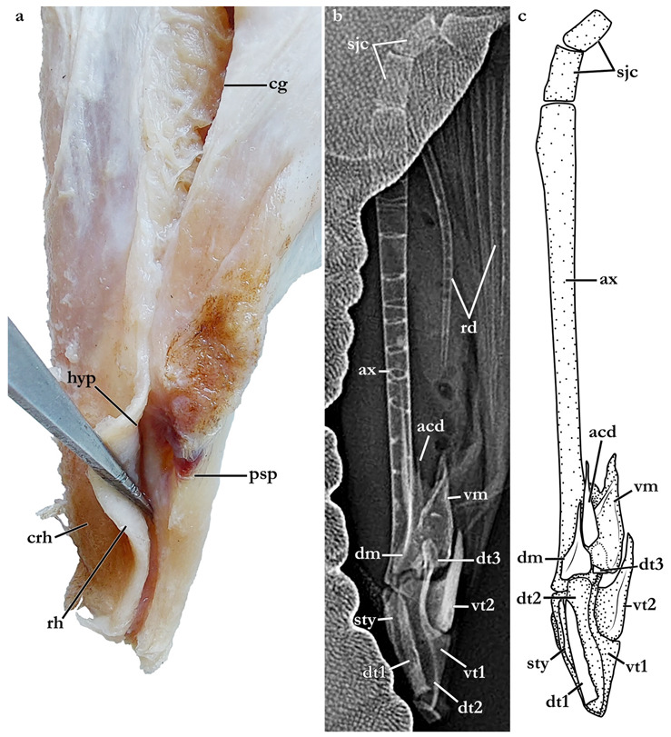
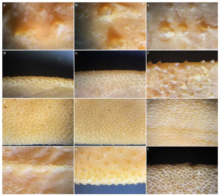
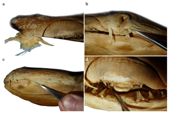
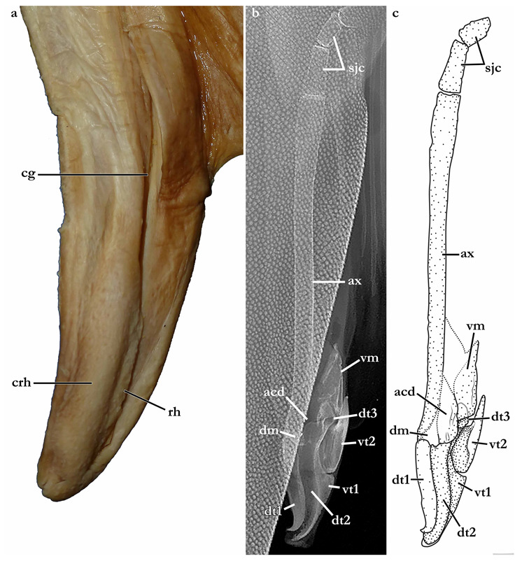
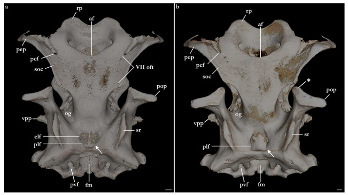
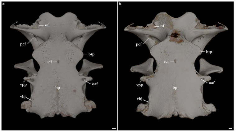
Sampling efforts on the Saya de Malha Bank (part of the Mascarene Plateau, western Indian Ocean) unveiled three unusual small juvenile angel shark specimens, that were a much paler color than the only known western Indian Ocean species, Squatina africana Regan, 1908. However, it took many years before further specimens, including adults of both sexes, and tissue samples were collected. The present manuscript contains a redescription of S. africana based on the holotype and additional material, as well as the formal description of the new species of Squatina. All specimens of the new species, hereafter referred to as Squatina leae sp. nov., were collected in the western Indian Ocean off southwestern India and on the Mascarene Plateau at depths of 100-500 m. The new species differs from S. africana in a number of characteristics including its coloration when fresh, smaller size at birth, size at maturity, and adult size, genetic composition, and distribution. Taxonomic characteristics include differences in the morphology of the pectoral skeleton and posterior nasal flap, denticle arrangement and morphology, vertebral counts, trunk width, pectoral-pelvic space, and clasper size. A key to the species of Squatina in the Indian Ocean is provided.

Keywords: CO1; Chondrichthyes; Elasmobranchii; NADH2; PCA; angel sharks; diversity; genetics; mCT scans; morphology; systematics; taxonomy.


Conflict of interest statement

The authors declare no conflict of interest. The funders had no role in the design of the study; in the collection, analyses, or interpretation of data; in the writing of the manuscript; or in the decision to publish the results.

Figures

Figure 18
Figure 18
Principal component analyses using standardized measurements of all specimens of Squatina leae sp. nov. and S. africana. (a) Scree plot demonstrating the percentage of the variance explained by each PC axis. (b) Morphospace plotted on PC1 in the x axis, PC2 in the y axis, and specimens of each species assigned to a separate group. (c) Morphospace plotted on PC1 and PC2, assigning groups to different stages of maturity across both species. (d). Morphospace plotted on PC1 and PC2, assigning groups to different sexes in specimens of both species. (e) Morphospace plotted on PC1 and PC2 with labels of each individual specimen plotted over the values of the variance of each measurement. (f) Values of variance of each morphological measurement plotted on PC1 and PC2. Color gradient represents the percentage of the variance explained by each morphological measurement.
Figure 1
Figure 1
Squatina leae sp. nov., holotype, CMFRI GA. 15.2.5.4, adult male, 671 mm TL, in (a) dorsolateral, (b) dorsal, and (c) ventral views in fresh condition. Photographs kindly provided by P. U. Zacharia (ICAR-CMFRI). Scale bar: 5 cm.
Figure 2
Figure 2
Squatina leae sp. nov., holotype, CMFRI GA. 15.2.5.4, adult male, 671 mm TL, head in (a) dorsal and (b) ventral views, (c) clasper region in dorsal view, (d) anterior pectoral-fin margin in dorsofrontal view, (e) dorsal fins in dorsal view, and (f) caudal fin in dorsolateral view. Photographs (ad,f) kindly provided by P. U. Zacharia (ICAR-CMFRI) show the holotype in fresh condition, photograph (e) shows the holotype in preserved condition.
Figure 3
Figure 3
Squatina leae sp. nov., (a,c,d) holotype, CMFRI GA. 15.2.5.4, adult male, 671 mm TL, (a) anterior nasal flaps in frontal view, (c) dermal fold in dorsolateral view, and (d) tooth rows in frontal view; (b) paratype, ZMH 26097, juvenile male, 282.6 mm TL, posterior nasal flap with additional barblet (indicated by white arrow) in frontal view. Photographs (a,c) kindly provided by P. U. Zacharia (ICAR-CMFRI) show the holotype in fresh condition, photographs (b,d) show the respective specimens in preserved condition.
Figure 4
Figure 4
Squatina leae sp. nov., holotype, CMFRI GA. 15.2.5.4, adult male, 671 mm TL, clasper external morphology (a) and skeleton (b,c); (a) detail of left clasper glans with rhipidion spread to show morphological details of the spermatic duct (image reversed), (b) radiograph and (c) drawing of the right clasper skeleton. Dashed lines in (c) represent margins of dorsal terminal three cartilage engulfed by ventral marginal cartilage. The photograph (a) was taken and kindly provided by K. R. Aju and Sreekumar (CMFRI Museum). Abbreviations: ax, axial cartilage; acd, accessory dorsal marginal cartilage; cg, clasper groove; crh, cover rhipidion; dm, dorsal marginal; dt1, dorsal terminal 1 cartilage; dt2, dorsal terminal 2 cartilage; dt3, dorsal terminal 3 cartilage; sty, end-style cartilage; psp, pseudopera; rh, rhipidion; sjc, stem-joint cartilage; rd, pelvic-fin radials; vm, ventral marginal; vt1, ventral terminal 1 cartilage; vt2, ventral terminal 2 cartilage.
Figure 5
Figure 5
Squatina leae sp. nov., paratype, ZMH 26097, juvenile male, 282.6 mm TL, tail in ventral view showing ventral precaudal ridge. Scale bar: 2 cm.
Figure 6
Figure 6
Squatina leae sp. nov., paratypes ZMH 26097, juvenile male, 298 mm TL fresh (in dorsal view) and ZMH 26098, juvenile male, 259 mm TL fresh (in ventral view) taken directly after catching. The photograph was taken and kindly provided by Matthias F. W. Stehmann. Scale bar: 5 cm.
Figure 7
Figure 7
Squatina leae sp. nov., unretained male specimen caught off Laccadive Islands, Southwest India, June 2017, in (a) dorsal and (b) ventral views taken directly after landing in Kochi, India. The photographs were kindly provided by Mr. Basheer.
Figure 8
Figure 8
Squatina leae sp. nov., holotype, CMFRI GA. 15.2.5.4, adult male, 671 mm TL, microscopic images of dermal denticles in (a) interorbital, (b,c) postorbital, (d) post-spiracle, (e) anterior pectoral-fin, and (f) predorsal regions.
Figure 9
Figure 9
Squatina leae sp. nov., paratype, ZMH 26097, juvenile male, 282.6 mm TL, microscopic images of dermal denticles in (a) preorbital, (b) postorbital, (c) predorsal, (d) anterior pectoral-fin, (e) anterior pelvic-fin, (f) anterior dorsal caudal-fin, (g) ventral anterior pectoral-fin, (h) ventral anterior pelvic-fin, (i) ventral midtail, (j) ventral tail before origin of caudal-fin, (k) ventral lateral caudal keel, and (l) ventral caudal-fin regions. Scale bar (al): 1 mm.
Figure 10
Figure 10
Map of the western Indian Ocean depicting the catch locations of the holotype (white star) and paratypes (white circles) of Squatina leae sp. nov., as well as the holotype (black star) and examined non-type specimens (black circles) of S. africana. The catch area of the 14 paratypes of S. leae sp. nov. at PMBC is indicated as a semitranslucent rectangle.
Figure 11
Figure 11
Squatina africana, (a,b) holotype, BMNH 1906.11.19.21, late subadult male, 837 mm TL, in (a) dorsal and (b) ventral views, as well as (c) non-type specimen, SAIAB 187381, adult male, 840 mm TL, in dorsal view. The photograph (c) was taken and kindly provided by Nkosinathi Mazungula (NRF-SAIAB).
Figure 12
Figure 12
Squatina africana, holotype, BMNH 1906.11.19.21, late subadult male, 837 mm TL, head in (a) dorsal and (b) ventral views, (c) clasper region in ventral view, (d) right pectoral fin in dorsal view, (e) dorsal fins in dorsolateral view (image reversed), and (f) caudal fin in dorsolateral view.
Figure 13
Figure 13
Squatina africana, holotype, BMNH 1906.11.19.21, late subadult male, 837 mm TL, (a) anterior nasal flaps in frontal view, (b) posterior nasal flap without additional barblet in frontal view, (c) dermal fold in lateral view, and (d) tooth rows in frontal view.
Figure 14
Figure 14
Squatina africana, (a) holotype, BMNH 1906.11.19.21, late subadult male, 837 mm TL, detail of right clasper glans to show external clasper morphology; (b,c) non-type specimen, SAIAB 187381, adult male, 840 mm TL, (b) radiograph and (c) drawing of the right clasper skeleton. Dashed lines in (c) represent margins of clasper cartilages that were not entirely visible in the radiograph. The radiograph (b) was taken and kindly provided by Nkosinathi Mazungula (NRF-SAIAB). Abbreviations: ax, axial cartilage; acd, accessory dorsal marginal cartilage; cg, clasper groove; crh, cover rhipidion; dm, dorsal marginal; dt1, dorsal terminal 1 cartilage; dt2, dorsal terminal 2 cartilage; dt3, dorsal terminal 3 cartilage; rh, rhipidion; sjc, stem-joint cartilage; rd, pelvic-fin radials; vm, ventral marginal; vt1, ventral terminal 1 cartilage; vt2, ventral terminal 2 cartilage.
Figure 15
Figure 15
Squatina africana, variation in color pattern: (a) ZMH 25561, juvenile male, 394 mm TL, (b) ZMH 26100, juvenile male, 446 mm TL, (c) ZMH 123064, juvenile female, 309 mm TL, (d) uncatalogued adult male, (e) ERB 0968, adult male, 810 mm TL, (f) ERB 0971, adult male, 820 mm TL (image reversed). The fresh photograph (d) was taken by Phil Heemstra and kindly provided by Elaine Heemstra (NRF-SAIAB), the fresh photographs (e,f) were taken and kindly provided by Frederik Mollen (ERB). Scale bars: 5 cm.
Figure 16
Figure 16
Squatina africana, holotype, BMNH 1906.11.19.21, late subadult male, 837 mm TL, dermal denticles in (a) right snout tip, (b) right postorbital, (c) right interspiracular, and (d) predorsal regions.
Figure 17
Figure 17
Squatina africana, non-type specimen, ZMH 123064, juvenile female, 309 mm TL, microscopic images of dermal denticles in (a) preorbital, (b) postorbital, (c) predorsal, (d) anterior pectoral-fin, (e) anterior pelvic-fin, (f) anterior dorsal caudal-fin, (g) anterior first-dorsal fin, (h) ventral anterior pectoral-fin, (i) ventral anterior pelvic-fin, (j) ventral tail before origin of caudal-fin, (k) ventral lateral caudal keel, and (l) ventral caudal-fin regions. Scale bar (al): 1 mm.
Figure 19
Figure 19
Squatina leae sp. nov., paratype, ZMH 26098, juvenile male, 249.6 mm TL; micro-computed tomography scans of the skull in (a) lateral and (b) dorsal views. Anterior to left in (a) and top in (b).
Figure 20
Figure 20
Squatina africana, ZMH 123064, juvenile female, 309 mm TL; micro-computed tomography scans of the skull: (a) dorsal view, (b) sagittal cross-section. Abbreviations: bh, basihyal; ch, ceratohyal; hb2-4, hypobranchials 2–4; Mc, Meckel’s cartilage; pef, pseudobranchial artery foramen; pq, palatoquadrate; I, olfactory bulb foramen; II, optic nerve foramen; III, oculomotor nerve foramen; IV, trochlear nerve foramen; V, trigeminal nerve foramen; VI, abducens nerve foramen; VII, facialis nerve foramen; VIII, auditory nerve foramen; IX, glossopharyngeal nerve foramen; X, vagus nerve foramen. Anterior to top in (a) and left in (b).
Figure 21
Figure 21
Micro-computed tomography scans of the neurocranium of (a) Squatina leae sp. nov., paratype, ZMH 26098, juvenile male, 249.6 mm TL and (b) S. africana, ZMH 123064, juvenile female, 309 mm TL in dorsal view. Abbreviations: af, anterior fontanelle; elf, endolymphatic foramen; fm, foramen magnum; og, orbital groove; pcf, preorbital canal foramen; pef, pseudobranchial artery foramen; plf, perilymphatic foramen; pop, postorbital process; pvf, posterior vein foramen; rp, rostral projections; soc, supraorbital crest; sr, sphenopterotic ridge; VII oft, foramina for rami of superficial ophthalmic nerve. The arrows indicate the margins of the perilymphatic foramen, distinctive to each species of Squatina. Squatina leae sp. nov. has concave margins, whereas in S. africana, the margins are convex. The asterisk marks the anteromedial projection of the postorbital process, exclusive to S. africana. Anterior to top; scale bars: 2 mm.
Figure 22
Figure 22
Micro-computed tomography scans of the neurocranium of (a) Squatina leae sp. nov., paratype, ZMH 26098, juvenile male, 249.6 mm TL and (b) S. africana, ZMH 123064, juvenile female, 309 mm TL in ventral view. Abbreviations: bp, basal plate; btp, basitrabecular process; icf, internal carotid foramen; nf, nasal foramen; oaf, orbital artery foramen; pcf, preorbital canal foramen; vhj, ventral hyoid junction; vpp, ventral postorbital process. Anterior to top; scale bars: 2 mm.
Figure 23
Figure 23
Micro-computed tomography scans of the neurocranium of (a,c,e) Squatina leae sp. nov., paratype, ZMH 26098, juvenile male, 249.6 mm TL and (b,d,f) S. africana, ZMH 123064, juvenile female, 309 mm TL in lateral view. Abbreviations: btp, basitrabecular process; epf, external profundus foramen; hf, hyomandibular facet; jc, jugular canal; lc, lateral commissure; oec, otic external canal; onc, orbitonasal canal; pcf, preorbital canal foramen; pef, pseudobranchial artery foramen; pop, postorbital process; soc, supraorbital crest; sr, sphenopterotic ridge; II, optic nerve foramen; III, oculomotor nerve foramen; IV, trochlear nerve foramen; V, trigeminal nerve foramen; VI, abducens nerve foramen; VII, facialis nerve foramen. Facing left; scale bars (a,b): 2 mm, (c,d,f): 1 mm.
Figure 24
Figure 24
Micro-computed tomography scans of the neurocranium of (a) Squatina leae sp. nov., paratype, ZMH 26098, juvenile male, 249.6 mm TL and (b) S. africana, ZMH 123064, juvenile female, 309 mm TL in occipital view. Abbreviations: fm, foramen magnum; occ, occipital condyle; ohc, occipital hemicentrum; pvf, posterior vein foramen; sr, sphenopterotic ridge; IX, glossopharyngeal nerve foramen; X, vagus nerve foramen. Scale bars: 2 mm.
Figure 25
Figure 25
Micro-computed tomography scans of the neurocranium of (a) Squatina leae sp. nov., paratype, ZMH 26098, juvenile male, 249.6 mm TL and (b) S. africana, ZMH 123064, juvenile female, 309 mm TL in frontal view. Abbreviations: btp, basitrabecular process; cr, cranial roof; epf, external profundus foramen; jc, jugular canal; lc, lateral commissure; onc, orbitonasal canal; pcc, precerebral cavity; pop, postorbital process; rp, rostral projections; sr, sphenopterotic ridge; vpp, ventral postorbital process. Scale bars: 2 mm.
Figure 26
Figure 26
Squatina leae sp. nov., paratype, ZMH 26098, juvenile male, 249.6 mm TL; micro-computed tomography scans of (a) cranium and jaws in lateral view, (b) jaws in medial view, upper jaw in (c) dorsal and (d) ventral views, and lower jaw in (e) dorsal and (f) ventral views. Abbreviations: alc, anterior upper labial cartilage; llc, lower labial cartilage; Mc, Meckel’s cartilage; plc, posterior upper labial cartilage; pq, palatoquadrate; qp, quadrate process. Scale bars: 2 mm.
Figure 27
Figure 27
Squatina leae sp. nov., paratype, ZMH 26098, juvenile male, 249.6 mm TL; micro-computed tomography scans of hyoid arch in (a) dorsal, (b) ventral, (c) lateral, and (d) ventral views, basihyal in (e) dorsal and (f) ventral views, ceratohyal in (g) dorsal and (h) ventral views, and hyomandibula in (i) dorsal and (j) ventral views. Abbreviations: bh, basihyal; ch, ceratohyal; hyo, hyomandibula; lcb, lateral cartilage of basihyal; vpc, ventral process of ceratohyal. Scale bars (a,b): 4 mm, (cj): 2 mm.
Figure 28
Figure 28
Micro-computed tomography scans of the branchial arches of (ad) Squatina leae sp. nov., paratype, ZMH 26098, juvenile male, 249.6 mm TL and (e,f) S. africana, ZMH 123064, juvenile female, 309 mm TL. (a,b) Epi- and pharyngobranchials in (a) dorsal and (b) ventral views, (cf) basi-, cerato-, and hypobranchials in (c,e) dorsal and (d,f) ventral views. Abbreviations: bb1–bb3, basibranchials 1–3; cb1–5, ceratobranchials 1–5; ep1–5, epibranchials 1–5; hb1–4, hypobranchials 1–4; pb1–5, pharyngobranchials 1–5. Anterior to top; scale bars: 5 mm.
Figure 29
Figure 29
Squatina leae sp. nov., paratype, ZMH 26098, juvenile male, 249.6 mm TL; micro-computed tomography scans of (a) pectoral fin and girdle in dorsal view and (b) pectoral girdle in ventral view. Abbreviations: co, coracoid bar; df, diazonal foramen; mes, mesopterygium; met, metapterygium; mtc, metacondyle; prc + msc, condyle for propterygium and mesopterygium; pro, propterygium; rd, radials; scl, scapula; ssc, suprascapula. Scale bars: 10 mm.
Figure 30
Figure 30
Micro-computed tomography scans of the pectoral girdle of (a,c) Squatina leae sp. nov., paratype, ZMH 26098, juvenile male, 249.6 mm TL and (b,d) S. africana, ZMH 123064, juvenile female, 309 mm TL in (a,b) frontal and (c,d) lateral views. Abbreviations: co, coracoid bar; df, diazonal foramen; ldf, lateral diazonal foramen; mtc, metacondyle; prc + msc, condyle for propterygium and mesopterygium; scl, scapula; ssc, suprascapula. Scale bars (a): 10 mm, (c,d): 5 mm.
Figure 31
Figure 31
Micro-computed tomography scans (a,b,d,e) and drawings (c,f) of the anterior pectoral skeleton of (a,b) Squatina leae sp. nov., paratype, ZMH 26098, juvenile male, 249.6 mm TL, (c) S. leae sp. nov., holotype, CMFRI GA. 15.2.5.4, adult male, 671 mm TL, (d,e) S. africana, ZMH 123064, juvenile female, 309 mm TL, (f) S. africana, holotype, BMNH 1906.11.19.21, late subadult male, 837 mm TL. The numbers in (c,f) indicate the number of radials articulating with the propterygium. Abbreviations: mes, mesopterygium; pro, propterygium; rd, radials.
Figure 32
Figure 32
Squatina leae sp. nov., paratype, ZMH 26098, juvenile male, 249.6 mm TL; micro-computed tomography scans of (a) pelvic fin in ventral view, as well as pelvic girdle in (b) dorsal and (c) ventral views. Abbreviations: ax, axial cartilage; bp, basipterygium; cbp, condyle for basipterygium; cfr, condyle for first enlarged pelvic radial; erd, enlarged radial; fbp, facet for basipterygium; lpp, lateral prepelvic process; of, obturator foramina; pb, pubioischiadic bar; rd, radials; sjc, stem-joint cartilages. Scale bars: 5 mm.
Figure 33
Figure 33
Maximum Likelihood Tree inferred from aligned CO1 sequences based on a General Time-Reversible Model with accommodation for both rate variation among sites and invariant sites. Parameter values estimated from the data. Type specimens noted where appropriate.
Figure 34
Figure 34
Maximum Likelihood Tree of phylogenetic relationships inferred from aligned NADH2 sequences based on a General Time-Reversible Model, with accommodation for both rate variation among sites and invariant sites. Parameter values were estimated from the data. Inferred relationships show strong geographical association. Type specimens noted where appropriate.

References

    1. Ebert D.A., Fowler S., Dando M. Sharks of the World: A Complete Guide. Princeton University Press; Princeton, NJ, USA: 2021.
    1. Claeson K.M., Hilger A. Morphology of the anterior vertebral region in elasmobranchs: Special focus, Squatiniformes. Foss. Rec. 2011;14:129–140. doi: 10.1002/mmng.201100003. - DOI
    1. Naylor G.J.P., Ryburn J.A., Fedrigo O., López J. Phylogenetic Relationships among the Major Lineages of Modern Elasmobranchs. In: Hamlett W.C., editor. Reprodutive Biology and Phylogeny of Chondrichthyes-Sharks, Batoids and Chimaeras. Science Publishers, Inc.; Enfield, UK: 2005. pp. 1–25.
    1. Naylor G.J.P., Caira J.N., Jensen K., Rosana K.A.M., White W.T., Last P.R. A DNA sequence–based approach to the identification of shark and ray species and its implications for global elasmobranch diversity and parasitology. Bull. Am. Mus. Nat. Hist. 2012;367:1–262. doi: 10.1206/754.1. - DOI
    1. Naylor G., Caira J., Jensen K., Rosana K., Straube N., Lakner C. Elasmobranch Phylogeny: A Mitochondrial Estimate Based on 595 Species. In: Carrier J.C., Musick J.A., Heithaus M.R., editors. Biology of Sharks and Their Relatives. CRC Press; Boca Raton, FL, USA: 2012. pp. 31–56.

LinkOut - more resources