Review
. 2023 Jul 14;12(7):1008.
doi: 10.3390/biology12071008.

Proline Isomerization: From the Chemistry and Biology to Therapeutic Opportunities

Affiliations
Review

Proline Isomerization: From the Chemistry and Biology to Therapeutic Opportunities

Deepti Gurung et al. Biology (Basel). .

Abstract

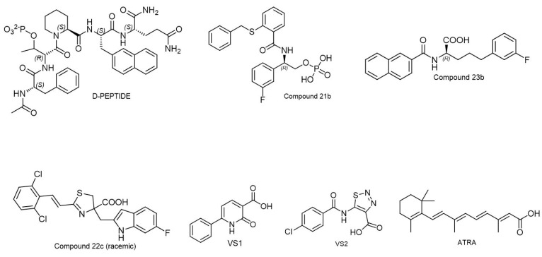
Proline isomerization, the process of interconversion between the cis- and trans-forms of proline, is an important and unique post-translational modification that can affect protein folding and conformations, and ultimately regulate protein functions and biological pathways. Although impactful, the importance and prevalence of proline isomerization as a regulation mechanism in biological systems have not been fully understood or recognized. Aiming to fill gaps and bring new awareness, we attempt to provide a wholistic review on proline isomerization that firstly covers what proline isomerization is and the basic chemistry behind it. In this section, we vividly show that the cause of the unique ability of proline to adopt both cis- and trans-conformations in significant abundance is rooted from the steric hindrance of these two forms being similar, which is different from that in linear residues. We then discuss how proline isomerization was discovered historically followed by an introduction to all three types of proline isomerases and how proline isomerization plays a role in various cellular responses, such as cell cycle regulation, DNA damage repair, T-cell activation, and ion channel gating. We then explore various human diseases that have been linked to the dysregulation of proline isomerization. Finally, we wrap up with the current stage of various inhibitors developed to target proline isomerases as a strategy for therapeutic development.

Keywords: 5-hydroxytryptamine type 3 (5-HT3); Alzheimer’s disease (AD); Ataxia telangiectasia and Rad3-related (ATR); FK506 binding protein (FKBP); Interleukin-2 inducible T-cell kinase (Itk); Parkinson’s disease (PD); Protein Interactor with NIMA1 (Pin1); autoimmune disease; cancer; cyclophilin; cyclosporin; hepatitis B virus (HBV); hepatitis C virus (HCV); human immunodeficiency virus 1 (HIV-1); infectious disease; multiple sclerosis (MS); neurodegenerative disease; parvulin; post-translation modifications; proline isomerase; proline isomerization; sanglifehrin; systemic lupus erythematosus (SLE).


Conflict of interest statement

The authors declare no conflict of interest.

Figures

Figure 1
Figure 1
Schematic diagram of protein peptide and the three torsion angles phi (Φ), psi (φ) and omega (ω) that define the conformation of protein backbone. The phi angle is around the -N-Cα- bond; the psi angle is around the -Cα-C- bond and the omega angle is around the -C-N- bond, which is also referred to as the peptide bond. Two sets of phi, psi and omega angles are labeled for residue at position m-1 and m in blue and green, respectively. (A). Peptide that has linear residues at position m-1, m and m+1. (B). Peptide that has a proline at position m. The freedom of the phi angle of proline is restricted due to the ring structure of proline.
Figure 2
Figure 2
Schematic diagram of trans- and cis-conformations around the peptidyl bond that connects residues m and m-1. To aid visualization, atoms around this peptidyl bond are depicted by spheres according to their sizes although not strictly proportionally. All spheres are in pink except for the two Cα atoms that are painted in green for easy identification. (A). Trans-conformation around the peptidyl bond connecting two linear residues. In this conformation, Cαm-1 and Cαm along with their connected linear R groups are dispersed on two sides of the peptidyl bond and therefore this conformation is energetically favorable. (B). Cis-conformation around the peptidyl bond connecting two linear residues. In this conformation, Cαm-1 and Cαm along with their connected linear R groups are concentrated on one side of the peptidyl bond, leading to crowdedness, or even clashing between atoms. Therefore, this conformation is energetically unfavorable. (C). Trans-conformation around the peptidyl bond connecting a linear residue with a proline. In this conformation, although Cαm-1 and Cαm are located on different sides of the peptidyl bond, due to the cyclic arrangement of atoms on proline, the Cδ atom replaces the hydrogen atom and wraps back to the backbone to be close to Cαm-1 and its connected Rm-1 group. This leads to the atoms being not as dispersed as in scenario (A). (D). Cis-conformation around the peptidyl bond connecting a linear residue with a proline. In this conformation, although Cαm-1 and Cαm are located on the same side of the peptidyl bond, because the cyclic arrangement of proline loops the side chain atoms back to the backbone, atoms are not as concentrated on one side as seen in scenario (B). As a result, the energy difference between the trans- and cis-conformation around a peptidyl-prolyl bond is much smaller than that of two linear residues.
Figure 3
Figure 3
Overall structures of all three types of PPIases. Ribbon representation is colored by secondary structure with yellow, magenta, and blue for helices, sheets, and loops, respectively. (A). Overall structure of cyclophilin (PDB code: 1MF8 [53]). (B). Overall structure of FKBP12 (PDB code: 1FKJ [54]). The ligand, FK506, is also displayed in ball-and-stick representation. (C). Overall structure of Pin1 (PDB code: 2ITK [55]). The PPIase domain is colored by secondary structure while the WW domain is shown by dark blue. The dotted line indicates some missing residues connecting the two domains. All three images are generated by UCSF Chimera [56].
Figure 4
Figure 4
(A). Schematic diagram to show how ATR is regulated by proline isomerization to switch between its dual roles in modulating cell death and DNA damage checkpoint signaling. (B). NMR structure of Cis- and trans-proline at position 287 switches the conformation of the Itk SH2 domain. Cis- and trans-conformations are colored in pink (PDB code: 1LUK [89]) and blue (PDB code: 1LUN [89]), respectively. Proline 287 and its preceding residue Asn286 are shown in ball and stick by red and dark blue in cis- and trans-conformation, respectively. The image is generated by using UCSF Chimera [56].
Figure 5
Figure 5
An illustration showing the impact of PPIase in four prominent human diseases. (A). Autoimmune disease. In multiple sclerosis, CypD plays a crucial role by binding and regulating mPTP. This interaction leads to the influx of small molecules depicted by green or orange spheres into the cells, causing mitochondrial swelling and rupture, cytochrome C release, and ultimately cell death. (B). Cancer. FKBP52 serves as the co-chaperone of HSP90 and interacts with steroid hormone receptors, including the androgen receptor (AR), glucocorticoid receptor (GR), and estrogen receptor (ER). This interaction enhances the transcriptional activity of the respective receptors, leading to increased cell proliferation, as observed in breast and prostate cancer. (C). Infectious disease. CypA plays a pivotal role in the life cycle of the HIV-1 virus by catalyzing proline isomerization at the Gly89-Pro90 site of the viral capsid, resulting in its conformational transition that is vital for capsid uncoating, viral genome release, and ultimately promoting viral replication. (D). Neurodegenerative disease. Pin1 plays a significant role in the pathogenesis of Alzheimer’s disease and Parkinson’s disease. In the case of Alzheimer’s disease, Pin1 catalyzes the isomerization of APP and tau from trans to cis, leading to the formation of amyloid plaques and neurofibrillary tangles, respectively, which are distinct hallmarks of Alzheimer’s disease. In the case of Parkinson’s disease, Pin1 interacts with phosphorylated synphilin-1, forming a stable complex with α-synuclein protein and promoting the formation of Lewy bodies, a characteristic feature of Parkinson’s disease. This figure is created using Inkscape (Version 1.2). The RNA icon is created by Servier (Source: Servier, https://smart.servier.com/ (accessed on 1 May 2023)) and is licensed under CC-BY 3.0 Unported (License: CC-BY 3.0 Unported, https://creativecommons.org/licenses/by/3.0/ (accessed on 1 May 2023)).
Figure 6
Figure 6
Chemical structures of cyclophilin inhibitors. (A). Molecular skeleton of cyclosporin. (B) Molecular skeleton of sanglifehrin A. Reference for cyclophilin inhibitors and their corresponding substitutes are shown on the right. Compounds are numbered and shown in bold. Structures of substitutes at different R positions for different compound number are given at the top. Compound names corresponding to compound number are shown in the table at the bottom. The structures are drawn using ChemDraw (Version 22.2.0).
Figure 7
Figure 7
Chemical structures of FKBP inhibitors. Rapamycin and FK506, the two FDA-approved natural ligands for immunosuppression, carry the pipecolate moiety critical for FKBP binding. The two GPI-derivatives carry a similar chemical group to the pipecolate moiety. SaFit1 and SaFit2 are the two inhibitors found to be selective to the FKBP51 isoform. The structures are drawn using ChemDraw (Version 22.2.0).
Figure 8
Figure 8
Chemical structures of Pin1 covalent inhibitors. Juglone, the first Pin1 covalent inhibitor, is a natural and simple compound with limited specificity to Pin1. KPT-6566, a derivative of Juglone with added structural complexity, shows higher specificity to Pin1 than Juglone. (S)-2 and BJP-06-005-3 are covalent inhibitors that do not share structural similarity with Juglone. ZL-Pin13 and Sulfopin, also distinct from Juglone, are two newly discovered covalent inhibitors with high selectivity and potency against Pin1. The structures are drawn using ChemDraw (Version 22.2.0).
Figure 9
Figure 9
Chemical structures of Pin1 non-covalent inhibitors. D-PEPTIDE is a non-natural peptide. Compound 21b, Compound 23b, and Compound 22c (racemic) developed by Pfizer have diverse structures with high potency inhibiting Pin1 activity in vitro. VS1 and VS2 are two newly discovered inhibitors with smaller molecular sizes. ATRA has been the only Pin1 inhibitor that has moved to clinical trials so far. The structures are drawn using ChemDraw (Version 22.2.0).

References

    1. Willstatter R. Synthesis of hygric acid. Berichte Dtsch. Chem. Ges. 1900;33:1160. doi: 10.1002/cber.190003301201. - DOI
    1. Fischer E. Hydrolysis of casein by means of hydrochloric acid. J. Chem. Soc. 1901;33:151–176.
    1. Brandts J.F., Halvorson H.R., Brennan M. Consideration of the Possibility that the slow step in protein denaturation reactions is due to cis-trans isomerism of proline residues. Biochemistry. 1975;14:4953–4963. doi: 10.1021/bi00693a026. - DOI - PubMed
    1. Schmid F.X., Baldwin R.L. Acid catalysis of the formation of the slow-folding species of RNase A: Evidence that the reaction is proline isomerization. Proc. Natl. Acad. Sci. USA. 1978;75:4764–4768. doi: 10.1073/pnas.75.10.4764. - DOI - PMC - PubMed
    1. Lin L.N., Brandts J.F. Role of cis-trans isomerism of the peptide bond in protease specificity. Kinetic studies on small proline-containing peptides and on polyproline. Biochemistry. 1979;18:5037–5042. doi: 10.1021/bi00590a002. - DOI - PubMed

LinkOut - more resources