Review
. 2023 Jul 12;12(14):1830.
doi: 10.3390/cells12141830.

A Role of PI3K/Akt Signaling in Oocyte Maturation and Early Embryo Development

Affiliations
Review

A Role of PI3K/Akt Signaling in Oocyte Maturation and Early Embryo Development

Jaroslav Kalous et al. Cells. .

Abstract

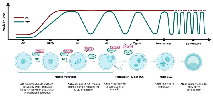
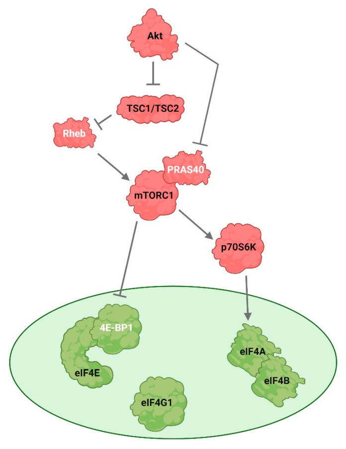
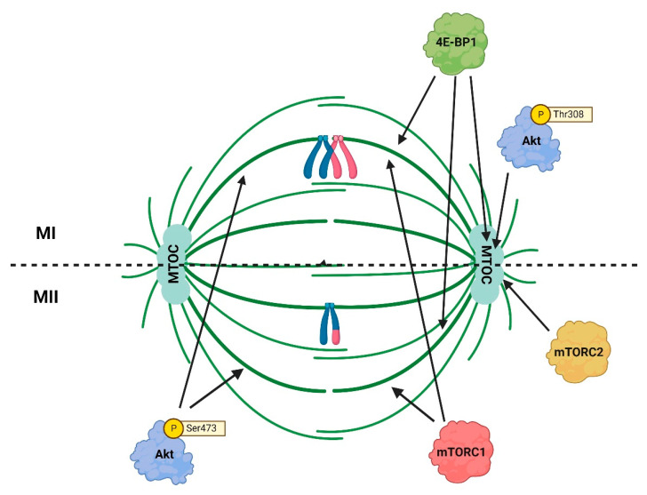
A serine/threonine-specific protein kinase B (PKB), also known as Akt, is a key factor in the phosphoinositide 3-kinase (PI3K)/Akt signaling pathway that regulates cell survival, metabolism and proliferation. Akt phosphorylates many downstream specific substrates, which subsequently control the nuclear envelope breakdown (NEBD), centrosome maturation, spindle assembly, chromosome segregation, and cytokinesis. In vertebrates, Akt is also an important player during oogenesis and preimplantation development. In the signaling pathways regulating mRNA translation, Akt is involved in the control of mammalian target of rapamycin complex 1 (mTORC1) and thereby regulates the activity of a translational repressor, the eukaryotic initiation factor 4E (eIF4E) binding protein 1 (4E-BP1). In this review, we summarize the functions of Akt in mitosis, meiosis and early embryonic development. Additionally, the role of Akt in the regulation of mRNA translation is addressed with respect to the significance of this process during early development.

Keywords: Akt kinase; early embryo; mRNA translation; mTORC1; meiosis; mitosis; oocyte; spindle.


Conflict of interest statement

The authors declare no conflict of interest.

Figures

Figure 1
Figure 1
Akt and maturation-promoting factor (MPF) activity during meiotic maturation, oocyte-to-embryo transition and early embryo development. During the first meiotic arrest at the germinal vesicle (GV) stage, Akt is inactivated. During germinal vesicle breakdown (GVBD), MPF and Akt activity increases. At meiosis I (MI) stage, Akt activity is remains stable and activity of MPF reaches maximum. At the MI/meiosis II (MII) transition, cyclin B is destructed by the anaphase promoting complex/cyclosome (APC/C), MPF activity is reduced, and the first polar body is extruded. The second metaphase plate is formed at the MII stage, and meiosis is stopped in the second meiotic arrest. Akt activity is maintained after the first polar body extrusion, MPF activity is restored to the MI levels, and the oocyte awaits fertilization. After fertilization, MPF activity is downregulated, Akt activity persists in the zygote, when the second polar body is extruded, and both male and female pronuclei are formed. MPF activity is restored before the first mitotic division. At the 2-cell embryonic stage, the first mitotic division is completed, Akt remains activated and MPF activity declines. Akt is involved in the regulation of zygotic genome activation (ZGA) which occurs in mouse embryo at the 2-cell stage. During the early embryo development, the Akt activity remains at high levels and MPF activity is cyclically downregulated and restored at each subsequent mitotic division.
Figure 2
Figure 2
Akt regulates mRNA translation. Akt directly activates mammalian target of rapamycin complex 1 (mTORC1) by phosphorylating and inactivating proline-rich Akt substrate 40 kDa (PRAS40), a protein that is associated with mTORC1. Further, Akt also activates mTORC1 indirectly by phosphorylating and inactivating the tuberous sclerosis complex 1 (TSC1) and tuberous sclerosis complex 2 (TCS2). TSC1/TSC2 functional complex acts as a key upstream negative regulator of mTORC1 activity. TSC2 has a role as a GTPase-activating protein (GAP) which inactivates an essential mTORC1 activator, the RAS homologue enriched in brain (Rheb). MTORC1 phosphorylates and activates the 70-kDa ribosomal protein S6 kinase (p70S6K), which phosphorylates the eukaryotic translation initiation factor 4B (eIF4B). The eukaryotic translation initiation factor 4E (eIF4E)-binding protein 1 (4E-BP1), phosphorylated and activated by mTORC1, is released from eIF4E and the assembly of the eIF4F complex is enabled. Factors involved in cap-dependent translation initiation are depicted in green, the upstream factors in red. Stimulatory modification is depicted as an arrow, inhibitory modification as a blunt end line.
Figure 3
Figure 3
Co-localization of Akt with 4E/BP1 and mTORC1/mTORC2 at the meiosis I (MI) and meiosis II (MII) spindles. The p473-Akt and mTORC1 are distributed along microtubules, the p308-Akt and mTORC2 are localized at spindle poles where microtubule-organizing centers (MTOCs) are positioned. 4E-BP1 is detected along microtubules and at spindle poles.

References

    1. Tanaka M., Kihara M., Hennebold J.D., Eppig J.J., Viveiros M.M., Emery B.R., Carrell D.T., Kirkman N.J., Meczekalski B., Zhou J., et al. H1FOO Is Coupled to the Initiation of Oocytic Growth. Biol. Reprod. 2005;72:135–142. doi: 10.1095/biolreprod.104.032474. - DOI - PubMed
    1. Alizadeh Z., Kageyama S.-I., Aoki F. Degradation of maternal mRNA in mouse embryos: Selective degradation of specific mRNAs after fertilization. Mol. Reprod. Dev. 2005;72:281–290. doi: 10.1002/mrd.20340. - DOI - PubMed
    1. Richter J.D., Lasko P. Translational Control in Oocyte Development. Cold Spring Harb. Perspect. Biol. 2011;3:a002758. doi: 10.1101/cshperspect.a002758. - DOI - PMC - PubMed
    1. Bianchi E., Sette C. Post-Transcriptional Control of Gene Expression in Mouse Early Embryo Development: A View from the Tip of the Iceberg. Genes. 2011;2:345–359. doi: 10.3390/genes2020345. - DOI - PMC - PubMed
    1. Manning B.D., Cantley L.C. AKT/PKB Signaling: Navigating Downstream. Cell. 2007;129:1261–1274. doi: 10.1016/j.cell.2007.06.009. - DOI - PMC - PubMed

Publication types

MeSH terms

Substances