Review
. 2023 Jul 13;12(14):1846.
doi: 10.3390/cells12141846.

Targeted Protein Degradation: Principles and Applications of the Proteasome

Affiliations
Review

Targeted Protein Degradation: Principles and Applications of the Proteasome

Yosup Kim et al. Cells. .

Abstract

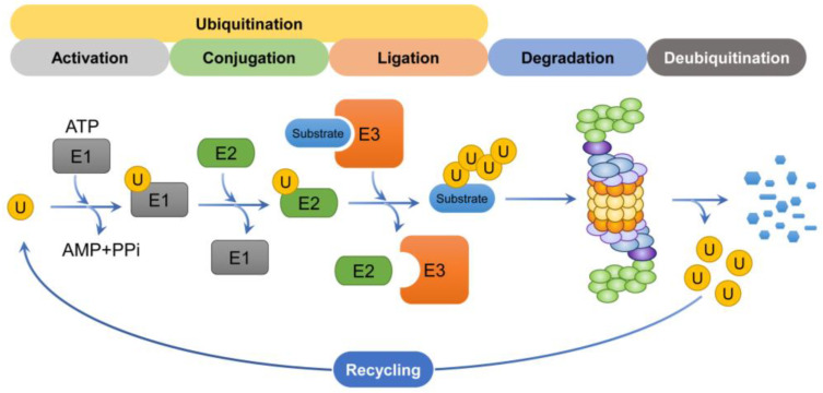
The proteasome is a multi-catalytic protease complex that is involved in protein quality control via three proteolytic activities (i.e., caspase-, trypsin-, and chymotrypsin-like activities). Most cellular proteins are selectively degraded by the proteasome via ubiquitination. Moreover, the ubiquitin-proteasome system is a critical process for maintaining protein homeostasis. Here, we briefly summarize the structure of the proteasome, its regulatory mechanisms, proteins that regulate proteasome activity, and alterations to proteasome activity found in diverse diseases, chemoresistant cells, and cancer stem cells. Finally, we describe potential therapeutic modalities that use the ubiquitin-proteasome system.

Keywords: E3 ubiquitin ligase; PROTAC; proteasome; targeted protein degradation; ubiquitin.


Conflict of interest statement

The authors declare no conflict of interest.

Figures

Figure 1
Figure 1
Schematic representation of the type and function of ubiquitin chains. Ubiquitin forms an isopeptide bond with substrate side-chain lysine (K), serine (S), threonine (T), and cysteine (C) residues through its C-terminal glycine residue 76 (G76). These chains, through mono-, multi-, and polyubiquitination, participate in various cellular signaling pathways. The representative sites and functions of the polyubiquitination chain are as follows (right).
Figure 2
Figure 2
The assembly of the 20S proteasome and two parts of the 19S regulatory particle, the lid and base subcomplexes. (A) Schematic diagram of the 20S proteasome. For synthesis of the 20S proteasome, the α-ring is formed first, followed by the formation of the β-subunit, thereby forming the β ring. Of the β-subunits, the catalytically active subunits that directly degrade are β1, β2, and β5. (B) Schematic diagram of the lid subcomplex. Rpn12 is an important subunit that binds to LP2 (i.e., module1 + LP3) to form the lid subcomplex. This is involved in base binding via Rpn10. (C) Schematic diagram of the base subcomplex. The basic subcomplex directly linked to 20S proteasome is complexed by various chaperone proteins. The lid subcomplex and base subcomplex are collectively referred to as 19S RPs.
Figure 3
Figure 3
Schematic representation of proteasome structure. Rpn10 acts as a hinge between the 20S proteasome and the 19S regulatory particle. Rpn10 is also involved in the formation of the 26S proteasome. As a result, one or two 19S regulatory particles associate with the 20S proteasome to form the 26S proteasome.
Figure 4
Figure 4
Schematic representation of the degradation process. The ubiquitin mediates the K48-linked ubiquitination of substrates through a cascade involving E1, E2, and E3 enzymes. Ubiquitin chains are recognized by the 19S regulatory particle and subsequently degraded by the 26S proteasome. The ubiquitin molecules that participated in the degradation process are then recycled to participate in substrate degradation again.
Figure 5
Figure 5
Mechanisms of action of PROTAC, molecular glues, and hydrophobic tagging. A variety of degraders are being developed for TPD. (A) PROTAC involves a protein of interest (POI) ligand and an E3 ligand that are linked by a linker. When POI and E3 ubiquitin ligase bind to PROTAC, the ubiquitin of E2 is sequentially attached to POI, thereby inducing ubiquitination. Ubiquitinated POIs are eventually degraded by the proteasome. (B) Molecular glues use a mechanism similar to PROTAC. They induce molecular proximity between a POI and E3 ubiquitin ligase, resulting in the sequential attachment of ubiquitin to the POI, which triggers its ubiquitination and degradation. (C) Hydrophobic tagging combines POIs to mimic hydrophobic and partially disordered forms of POIs. Recognition of chaperones at these sites then leads to chaperone-mediated proteasomal degradation.

References

    1. Apel K., Hirt H. Reactive oxygen species: Metabolism, oxidative stress, and signal transduction. Annu. Rev. Plant Biol. 2004;55:373–399. doi: 10.1146/annurev.arplant.55.031903.141701. - DOI - PubMed
    1. D’Autreaux B., Toledano M.B. ROS as signalling molecules: Mechanisms that generate specificity in ROS homeostasis. Nat. Rev. Mol. Cell Biol. 2007;8:813–824. doi: 10.1038/nrm2256. - DOI - PubMed
    1. Lobo V., Patil A., Phatak A., Chandra N. Free radicals, antioxidants and functional foods: Impact on human health. Pharmacogn. Rev. 2010;4:118–126. doi: 10.4103/0973-7847.70902. - DOI - PMC - PubMed
    1. Michalska P., Leon R. When It Comes to an End: Oxidative Stress Crosstalk with Protein Aggregation and Neuroinflammation Induce Neurodegeneration. Antioxidants. 2020;9:740. doi: 10.3390/antiox9080740. - DOI - PMC - PubMed
    1. Barral J.M., Broadley S.A., Schaffar G., Hartl F.U. Roles of molecular chaperones in protein misfolding diseases. Semin. Cell Dev. Biol. 2004;15:17–29. doi: 10.1016/j.semcdb.2003.12.010. - DOI - PubMed

Publication types

LinkOut - more resources