. 2023 Jul 18;12(14):1883.
doi: 10.3390/cells12141883.

Pyrroline-5-Carboxylate Reductase-2 Promotes Colorectal Carcinogenesis by Modulating Microtubule-Associated Serine/Threonine Kinase-like/Wnt/β-Catenin Signaling

Affiliations

Pyrroline-5-Carboxylate Reductase-2 Promotes Colorectal Carcinogenesis by Modulating Microtubule-Associated Serine/Threonine Kinase-like/Wnt/β-Catenin Signaling

Raju Lama Tamang et al. Cells. .

Abstract

Background: Despite significant progress in clinical management, colorectal cancer (CRC) remains the third most common cause of cancer-related deaths. A positive association between PYCR2 (pyrroline-5-carboxylate reductase-2), a terminal enzyme of proline metabolism, and CRC aggressiveness was recently reported. However, how PYCR2 promotes colon carcinogenesis remains ill understood.

Methods: A comprehensive analysis was performed using publicly available cancer databases and CRC patient cohorts. Proteomics and biochemical evaluations were performed along with genetic manipulations and in vivo tumor growth assays to gain a mechanistic understanding.

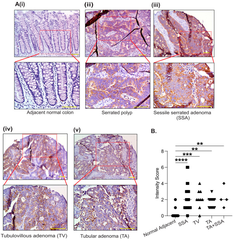
Results: PYCR2 expression was significantly upregulated in CRC and associated with poor patient survival, specifically among PYCR isoforms (PYCR1, 2, and 3). The genetic inhibition of PYCR2 inhibited the tumorigenic abilities of CRC cells and in vivo tumor growth. Coinciding with these observations was a significant decrease in cellular proline content. PYCR2 overexpression promoted the tumorigenic abilities of CRC cells. Proteomics (LC-MS/MS) analysis further demonstrated that PYCR2 loss of expression in CRC cells inhibits survival and cell cycle pathways. A subsequent biochemical analysis supported the causal role of PYCR2 in regulating CRC cell survival and the cell cycle, potentially by regulating the expression of MASTL, a cell-cycle-regulating protein upregulated in CRC. Further studies revealed that PYCR2 regulates Wnt/β-catenin-signaling in manners dependent on the expression of MASTL and the cancer stem cell niche.

Conclusions: PYCR2 promotes MASTL/Wnt/β-catenin signaling that, in turn, promotes cancer stem cell populations and, thus, colon carcinogenesis. Taken together, our data highlight the significance of PYCR2 as a novel therapeutic target for effectively treating aggressive colon cancer.

Keywords: MASTL; Wnt signaling; cancer progression; colorectal cancer (CRC); proline metabolism; proteomics; pyrroline 5 carboxylate reductases (PYCRs).


Conflict of interest statement

The authors declare no conflict of interest.

Figures

Figure 1
Figure 1
PYCR2 expression increases significantly in colon cancer. (AC) PYCR2 mRNA expression in Asian and European CRC cohorts (p = 0.000201 for Korean cohort, p = 2.003 × 10⁻¹¹ for French cohort, and p = 0.01577093 for Amsterdam cohort). (D) Analysis of PYCR2 protein expression in colorectal cancer patients in the CPTAC database (adjacent normal vs. primary tumor, p = 1.49 × 10−43). (E) PYCR2 protein expression in different stages of colorectal cancer (normal vs. stage 1, p = 6.5 × 10⁻⁵; normal vs. stage 2, p = 2.13 × 10⁻²²; normal vs. stage 3, p = 2.94 × 10⁻¹⁶; and normal vs. stage 4, p = 5.49 × 10⁻⁴). (F) Representative image showing PYCR2 expression in colon tumors of APCmin mice. (G) Representative images of immunohistochemical analysis of PYCR2 expression in colon adenoma and adenocarcinoma in comparison to normal adjacent human colon. (H) Quantitative analysis of PYCR2 expression in human colon polyps and CRC samples. **** p < 0.0001.
Figure 4
Figure 4
Inhibition of PYCR2 expression inhibits xenograft tumor growth and promotes apoptosis. (A) Schematics of in vivo studies using murine models of subcutaneous xenograft tumor growth and colonoscopy-guided cancer cell transplantation into the colon wall. (Bi) Representative images of the tumors isolated from athymic/nude mice injected with control or PYCR2-inhibited SW620 cells. (Bii,Biii) Statistical analysis showing % change in tumor volume (p = 0.0048) and fold change in tumor weight (p = 0.0019). (Ci) The analysis of the probability of mouse survival after colonoscopy-guided injection. (CiiCiv) Representative images of the quantification of the % of tumor development; respective images of colon tumors and tumor size quantification (p = 0.0014, control vs. PYCR2 KD). (Di) Representative H&E images of the tumors. (DiiDv) Representative images of IHC using anti-cleaved caspase-3 and p-H2AX antibodies in xenograft tumors and quantitative analysis (p = 0.0349 and p = 0.0018). (E) Immunoblot analysis for p-H2AX and cleaved PARP in HCT116 control and PYCR2-KD cells. (F,G) FACS analysis for early and late apoptosis in HCT116 control and PYCR2-KD cells and quantitative analysis (p = 0.0085). The representative figure has four quadrants where A = live cells, B = early apoptosis, C = late apoptosis, and D = necrosis. Data are presented as mean + SEM, and significance was determined using Student’s t-test and one-way ANOVA. * p < 0.05, ** p < 0.01, and *** p < 0.001.
Figure 2
Figure 2
PYCR2 protein expression is significantly upregulated in all histological types of CRC adenomas. (AiAv) Representative images of immunohistochemical analysis of PYCR2 expression in different types of colon adenomas and adjacent normal colon (TMA, N#109). (B) Scoring analysis of PYCR2 immunostaining intensity in normal adjacent colon vs. colon adenoma (p < 0.0001 for SSA, p = 0.002 for TV, p = 0.0015 for TA, and TA + SSA respectively). The data are presented as mean + SEM. Statistical significance was determined using one-way ANOVA and a post hoc Tukey’s test for pairwise comparison. **** p < 0.0001, *** p < 0.001, and ** p < 0.01.
Figure 3
Figure 3
Genetic manipulation of PYCR2 expression modulates oncogenic properties of CRC cells. (A) Western blot analysis of PYCR2 expression in different CRC cell lines. The IEC-6 cells served as normal intestinal epithelial cells. (Bi,Bii) Immunoblot analysis of control and genetically manipulated PYCR2 HCT116 and SW480 cells and densitometric analysis (p = 0.0021 and p = 0.015). (C,D) Representative immunoblot analysis and densitometric analysis of EpCAM, E-cadherin, and vimentin in control and PYCR2-KO HCT116 cells (p = 0.00012 for EpCAM and p = 0.021 and 0.019 for E-cadherin and vimentin). (E) Immunofluorescence staining images for EpCAM expression in control and PYCR2-KO HCT116 cells. (F) Cell proliferation assays using the HCT116 control and PYCR2-KD cells (p < 0.0001), (Gi,Gii) Soft agar assay using the HCT116 control and PYCR2-KD cells (p = 0.0257), (Hi,Hii) Cell migration assay using the HCT116 control and PYCR2-KD cells (p = 0.0355 at 48 h and p = 0.0048 at 72 h), (Ii,Iii) Cell invasion in HCT116 control and PYCR2-KD cells (p = 0.0017), and quantitative analysis. (J,K) Representative images of the immunoblot analysis of EpCAM, E-cadherin, and vimentin in control and PYCR2-overexpressing SW480 cells and densitometric evaluation (p = 0.031 for EpCAM and p = 0.0015 and 0.00029 for E-cadherin and vimentin). (L) Representative data for the effect of PYCR2 overexpression on cell proliferation (p = 0.00019). (Mi,Mii) Representative data for the cell invasion (p = 0.0024) in control and PYCR2-overexpressing SW480 cells. Data are presented as mean + SEM. Statistical significance was determined using Student’s t-test and one-way ANOVA. * p < 0.05, ** p < 0.01, *** p < 0.001, and **** p < 0.0001.
Figure 5
Figure 5
LC-MS/MS proteomics analysis to determine effects of PYCR2 loss of expression. (A) Schematics of the LC-MS/MS proteomics analysis. (B) Principal component analysis of the proteins differentially expressed in the control and PYCR2-KO HCT116 cells. (C,D) Analyses of the KEGG pathway and the GO biological function for differentially expressed proteins in PYCR2-KO versus control cells.
Figure 6
Figure 6
PYCR2 regulates cell survival pathways and cancer stem cell population. (A) Heatmap analysis of proteins involved in cell apoptosis and proliferation. (BG) Immunoblotting and densitometric analysis examining the expression of p-AKT and cyclin D1 in control and PYCR2-inhibited HCT116 and SW620 cells. (HJ) Immunoblotting and densitometric analysis examining the expression of p-AKT and cyclin D1 in control and PYCR2-overexpressing SW480 cells. (K) mRNA expression analysis for colonic CSC markers in HCT116 control cells and PYCR2-KD cells (p < 0.011 for CD133, and p = 0.00014 for CD44 and 0.9484 for Sox2 (ns). (L) Representative immunoblots for the analysis of colonic CSC markers in control and PYCR2-KO HCT116 cells. (Mi,Mii) Sphere-forming assay using HCT116 control cells and PYCR2-KD cells and quantitative analysis (p = 0.0018). (N) mRNA expression analysis of colonic CSC markers in control and PYCR2-overexpressing SW480 cells (p < 0.769 for CD133 (ns), 0.00156 for CD44, and p < 0.0001 for Sox2). (O) Representative immunoblots for the analysis of colonic CSC markers in control and SW480-PYCR2 cells. (PiPiii) Sphere-forming assay using control and SW480-PYCR2 cells and quantitative analysis (p = 0.0158 for number of spheres, and p = 0.00013 for size). Data are presented as mean + SEM. Statistical significance was determined using Student’s t-test and one-way ANOVA. ns = non-significant, ** p < 0.01, *** p < 0.001, and **** p < 0.0001.
Figure 7
Figure 7
PYCR2 regulates the cell cycle and modulates MASTL/Wnt/β-catenin signaling. (A,B) Representative images of the cell cycle analysis using the control and PYCR2-KO HCT116 cells showing cell cycle arrest in PYCR2-KO cells at the G2/M phase and the subsequent quantification of the % of cells arrested at the G2/M phase. (Ci,Cii) Representative images of immunoblots and densitometric analysis examining the effects of PYCR2 on MASTL expression in PYCR2-KO HCT116. (Di,Dii) Immunoblots and densitometric analysis for MASTL expression in control and SW480-PYCR2 cells. (Ei,Eii) Representative images of immunoblots and densitometric analysis examining the effects of PYCR2 on Wnt signaling (p-β catenin s552) using the control and PYCR2-KO HCT116. (F) TOP-flash luciferase-based analysis of control and PYCR2-KO HCT116 cells. (Gi,Gii) Effect of PYCR2 overexpression on Wnt signaling (p-β catenin s552) in control and SW480-PYCR2 cells followed by densitometric analysis. (H) TOP-flash activity analysis of control and SW480-PYCR2 cells. Data are presented as mean + SEM. Statistical significance was determined using Student’s t-test and one-way ANOVA. * p < 0.05, ** p < 0.01, *** p < 0.001, and **** p < 0.0001.
Figure 8
Figure 8
MASTL mediates CRC-promoting effects of PYCR2 expression. (AiAiii) Immunoblot analysis determining the effects of MASTL overexpression in PYCR2-KO HCT116 cells and densitometric analysis of MASTL and p-βcatenin s552 expression in control, PYCR2 KO, and MASTL overexpression in PYCR2 KO HCT116 cells. (BiBiii) Immunoblot analysis determining the effect of the GKI-an inhibitor on MASTL expression/activity in PYCR2-overexpressing SW480 cells. A densitometric analysis of MASTL and p-βcatenin s552 expression in control, PYCR2 overexpression, and MASTL-inhibited SW480 cells is also presented. (C,D) Cell proliferation assay of HCT116-KD and SW480-PYCR2 cells after MASTL overexpression and inhibition, respectively. (E) Schematics summarizing our findings on the regulatory role of PYCR2 in CRC progression caused by modulating MASTL/Wnt/β-catenin signaling. Data are presented as mean + SEM. Statistical significance was determined using Student’s t-test and one-way ANOVA. ns = non-significant, * p < 0.05, ** p < 0.01 and **** p < 0.0001.

References

    1. Siegel R.L., Miller K.D., Goding Sauer A., Fedewa S.A., Butterly L.F., Anderson J.C., Cercek A., Smith R.A., Jemal A. Colorectal cancer statistics, 2020. CA Cancer J. Clin. 2020;70:145–164. doi: 10.3322/caac.21601. - DOI - PubMed
    1. Qiu M., Hu J., Yang D., Cosgrove D.P., Xu R. Pattern of distant metastases in colorectal cancer: A SEER based study. Oncotarget. 2015;6:38658–38666. doi: 10.18632/oncotarget.6130. - DOI - PMC - PubMed
    1. Adorno-Cruz V., Kibria G., Liu X., Doherty M., Junk D.J., Guan D., Hubert C., Venere M., Mulkearns-Hubert E., Sinyuk M., et al. Cancer stem cells: Targeting the roots of cancer, seeds of metastasis, and sources of therapy resistance. Cancer Res. 2015;75:924–929. doi: 10.1158/0008-5472.CAN-14-3225. - DOI - PMC - PubMed
    1. Endo H., Owada S., Inagaki Y., Shida Y., Tatemichi M. Metabolic reprogramming sustains cancer cell survival following extracellular matrix detachment. Redox Biol. 2020;36:101643. doi: 10.1016/j.redox.2020.101643. - DOI - PMC - PubMed
    1. D’Aniello C., Patriarca E.J., Phang J.M., Minchiotti G. Proline Metabolism in Tumor Growth and Metastatic Progression. Front. Oncol. 2020;10:776. doi: 10.3389/fonc.2020.00776. - DOI - PMC - PubMed

Publication types

LinkOut - more resources