

An In Silico Analysis Reveals Sustained Upregulation of Neuroprotective Genes in the Post-Stroke Human Brain
- PMID: 37508918
- PMCID: PMC10377198
- DOI: 10.3390/brainsci13070986
An In Silico Analysis Reveals Sustained Upregulation of Neuroprotective Genes in the Post-Stroke Human Brain
Abstract
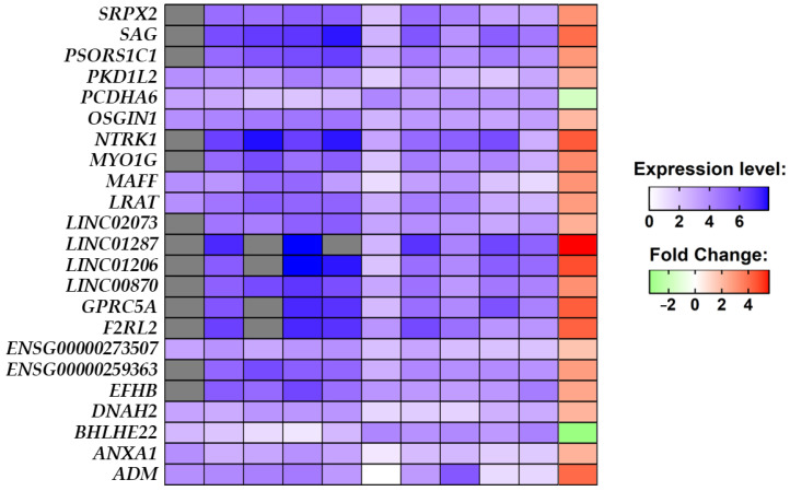
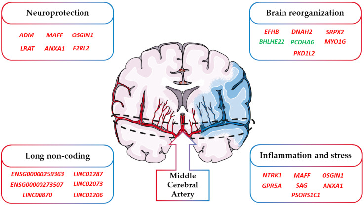
Ischemic stroke is a cerebrovascular disease caused by an interruption of blood flow to the brain, thus determining a lack of oxygen and nutrient supply. The ischemic event leads to the activation of several molecular signaling pathways involved in inflammation and the production of reactive oxygen species, causing irreversible neuronal damage. Several studies have focused on the acute phase of ischemic stroke. It is not clear if this traumatic event can influence some of the molecular processes in the affected area even years after the clinical event. In our study, we performed an in silico analysis using freely available raw data with the purpose of evaluating the transcriptomic state of post-mortem brain tissue. The samples were taken from non-fatal ischemic stroke patients, meaning that they suffered an ischemic stroke and lived for a period of about 2 years after the event. These samples were compared with healthy controls. The aim was to evaluate possible recovery processes useful to mitigating neuronal damage and the detrimental consequences of stroke. Our results highlighted differentially expressed genes codifying for proteins along with long non-coding genes with anti-inflammatory and anti-oxidant functions. This suggests that even after an amount of time from the ischemic insult, different neuroprotective mechanisms are activated to ameliorate brain conditions and repair post-stroke neuronal injury.
Keywords: human brain; in silico analysis; ischemic stroke; neuroinflammation; neuroprotection; oxidative stress; post-stroke; transcriptome.
Conflict of interest statement
The authors declare no conflict of interest. The funders had no role in the design of the study; in the collection, analysis, or interpretation of the data; in the writing of the manuscript; or in the decision to publish the results.
Figures
Similar articles
-
Emerging Roles of Sirtuins in Ischemic Stroke.Transl Stroke Res. 2017 Jun 27. doi: 10.1007/s12975-017-0544-4. Online ahead of print. Transl Stroke Res. 2017. PMID: 28656393 Review.
-
Computational systems pharmacology reveals an antiplatelet and neuroprotective mechanism of Deng-Zhan-Xi-Xin injection in the treatment of ischemic stroke.Pharmacol Res. 2019 Sep;147:104365. doi: 10.1016/j.phrs.2019.104365. Epub 2019 Jul 23. Pharmacol Res. 2019. PMID: 31348992
-
Neuron protection as a therapeutic target in acute ischemic stroke.Curr Top Med Chem. 2009;9(14):1317-34. doi: 10.2174/156802609789869646. Curr Top Med Chem. 2009. PMID: 19849659 Review.
-
Temporal expression profiling of DAMPs-related genes revealed the biphasic post-ischemic inflammation in the experimental stroke model.Mol Brain. 2020 Apr 7;13(1):57. doi: 10.1186/s13041-020-00598-1. Mol Brain. 2020. PMID: 32264906 Free PMC article.
-
MiRNA: Involvement of the MAPK Pathway in Ischemic Stroke. A Promising Therapeutic Target.Medicina (Kaunas). 2021 Oct 1;57(10):1053. doi: 10.3390/medicina57101053. Medicina (Kaunas). 2021. PMID: 34684090 Free PMC article. Review.
Cited by
-
STIM Proteins: The Gas and Brake of Calcium Entry in Neurons.Neurosci Bull. 2025 Feb;41(2):305-325. doi: 10.1007/s12264-024-01272-5. Epub 2024 Sep 12. Neurosci Bull. 2025. PMID: 39266936 Review.
References
Grants and funding
LinkOut - more resources
Full Text Sources
