Review
. 2023 Jul 14;13(7):1130.
doi: 10.3390/biom13071130.

A Frame-by-Frame Glance at Membrane Fusion Mechanisms: From Viral Infections to Fertilization

Affiliations
Review

A Frame-by-Frame Glance at Membrane Fusion Mechanisms: From Viral Infections to Fertilization

Farshad C Azimi et al. Biomolecules. .

Abstract

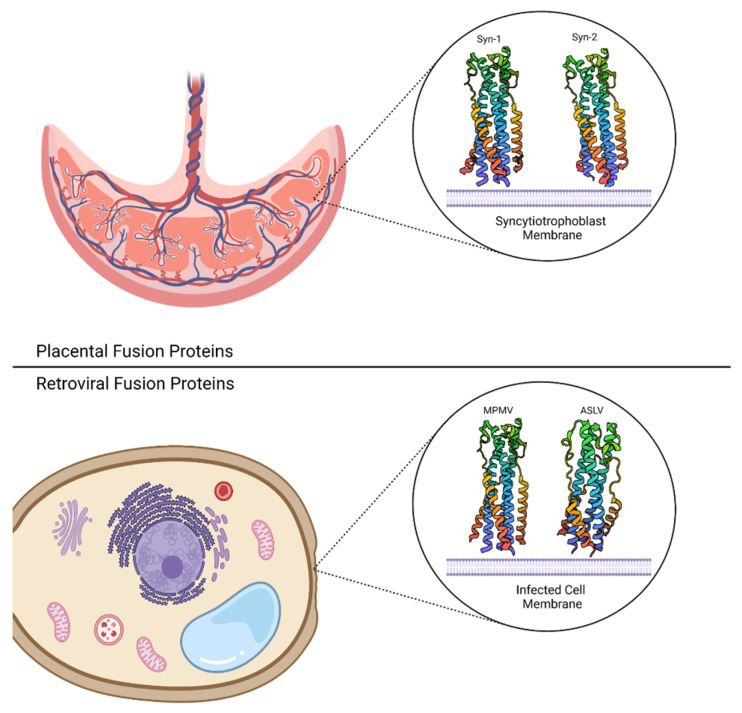
Viral entry and fertilization are distinct biological processes that share a common mechanism: membrane fusion. In viral entry, enveloped viruses attach to the host cell membrane, triggering a series of conformational changes in the viral fusion proteins. This results in the exposure of a hydrophobic fusion peptide, which inserts into the host membrane and brings the viral and host membranes into close proximity. Subsequent structural rearrangements in opposing membranes lead to their fusion. Similarly, membrane fusion occurs when gametes merge during the fertilization process, though the exact mechanism remains unclear. Structural biology has played a pivotal role in elucidating the molecular mechanisms underlying membrane fusion. High-resolution structures of the viral and fertilization fusion-related proteins have provided valuable insights into the conformational changes that occur during this process. Understanding these mechanisms at a molecular level is essential for the development of antiviral therapeutics and tools to influence fertility. In this review, we will highlight the biological importance of membrane fusion and how protein structures have helped visualize both common elements and subtle divergences in the mechanisms behind fusion; in addition, we will examine the new tools that recent advances in structural biology provide researchers interested in a frame-by-frame understanding of membrane fusion.

Keywords: cryo-electron microscopy; fertilization; fusion mechanism; membrane fusion; structural biology; viruses.


Conflict of interest statement

The authors declare no conflict of interest.

Figures

Figure 1
Figure 1
Membrane fusion mechanism. The process of membrane fusion, which is a crucial cellular mechanism involved in various biological processes, is divided into four distinct stages, each representing a key step in the fusion process. At the initial stage, two separate lipid bilayers are shown in close proximity to each other. The membranes are depicted as curved structures with different colors, highlighting their distinct identities. For membrane docking and recognition, the two opposing membranes undergo a series of molecular interactions to facilitate fusion. Proteins and other molecules aid in the recognition of specific fusion partners and ensure proper alignment of the membranes. The final stage showcases the actual fusion event between the membranes. The lipid bilayers seamlessly merge, resulting in the formation of a single continuous membrane structure. This fusion process enables the mixing of cellular contents and facilitates the exchange of molecules between the previously separate compartments. Created with BioRender.
Figure 2
Figure 2
Mechanisms of SNARE proteins in membrane fusion. The process begins with the docking of a vesicle to the target membrane. The vesicle and target membranes come into close proximity, facilitated via specific interactions between SNARE proteins (PDB ID 2C5J) present on both membranes. SNARE proteins on the vesicle membrane (v-SNAREs) and the target membrane (t-SNAREs) interact with each other, forming a highly stable and tightly coiled complex known as the SNARE complex. This complex consists of a four-helix bundle, with the v-SNARE and t-SNAREs contributing two helices each. The SNARE complex undergoes a process called “zippering”. This involves the progressive and tight association of the four helices, pulling the vesicle and target membranes closer together. The energy released during this zippering process helps overcome the repulsive forces between the membranes, leading to their fusion. As the SNARE complex completes its zippering, the lipid bilayers of the vesicle and target membranes merge, allowing the contents of the vesicle to be released into the target compartment. This process is accompanied by the mixing of transmembrane proteins and lipids, facilitating the exchange of molecules between the two compartments. After membrane fusion, the SNARE complex disassembles with the help of ATPase NSF (N-ethylmaleimide-sensitive factor) and its cofactor SNAPs (soluble NSF attachment proteins). This disassembly step prepares the SNARE proteins for subsequent rounds of membrane fusion. Created with BioRender.
Figure 3
Figure 3
Viral—cell membrane fusion mechanisms. (A) A generalized schematic of the viral fusion to cellular membranes. Homo-dimeric or homo-trimeric (and in some cases heterodimeric) pre-fusion complexes initiate attachment via cellular receptors, such as cellular membrane proteins or surface sugar moieties. Following a fusion trigger event (full receptor engagement, acidic pH, or both), the pre-fusion complex undergoes conformational changes that result in the insertion of the hydrophobic fusion peptide or a series of fusion loops into the cellular membrane, forming a high-energy pre-hairpin trimer intermediate. The pre-hairpin complex undergoes stabilization via the collapsing of its N- and C-termini, hence pulling the two membranes towards one another, resulting in the mixing of viral and cellular membranes and the formation of the fusion pore. (B) Representative structures of class I, II, and III viral fusion complexes in pre-fusion and post-fusion states. The pre-fusion complexes of class I and III are trimeric, while class II complexes are either homo-dimeric (shown here) or hetero-dimeric in nature and run parallel to the viral membrane (heterodimers can, in some cases, trimerize in the mature form). All classes possess trimeric post-fusion conformations. For a side-by-side comparison of the fusion complexes of the three classes, see Table 1.
Figure 4
Figure 4
Placental and retroviral fusion protein six-helix bundles (6HBs). The Syncytin-1 (PDB ID: 6RX1) and Syncytin-2 (PDB ID: 6RX3) 6HBs are oriented relative to the placental syncytiotrophoblast membrane (top), while the Mason-Pfizer Monkey Virus (PDB ID: 4JF3) and Avian Rous Sarcoma Virus (PDB ID: 4JPR) 6HBs are shown relative to the infected cell membrane (bottom). Both placental and retroviral proteins form the canonical post-fusion 6HB conformation following receptor recognition and/or pH acidification. Created with Biorender.
Figure 5
Figure 5
Membrane fusion as part of the Plasmodium life cycle. The sexual reproductive portion of the Plasmodium life cycle takes place in the mosquito vector, where haploid gametes merge to form diploid zygotes. This process requires the fusion protein HAP2/GCS1 to undergo a conformational change from a pre-fusion state (likely monomeric) to a post-fusion trimer; in the process, the relative orientations of domains 1 (red), 2 (yellow), and 3 (blue) also change. Since no solved structures of Plasmodium HAP2/GCS1 exist, representatives of the pre-fusion and post-fusion state are taken from Cyanidioschyzon merolae (PDB: 7S0K) and Chlamydomonas reinhardtii (PDB: 6E18), respectively. Therapeutic inhibitors targeting either the insertion of hydrophobic fusion loops into the membrane or the trimerization event could potentially prevent the formation of zygotes, thereby preventing the formation of haploid sporozoites that instigate further infections. Created with BioRender.
Figure 6
Figure 6
Key proteins involved in sperm-egg fusion. JUNO (purple) and MAIA (light green) are localized in the egg’s membrane in females. In males, IZUMO1 (blue), SPACA6 (yellow), TMEM95 (light blue), and FIMP (pink) are in the equatorial regions of the sperm. DCST1 (red) and DCST2 (dark grey) are transmembrane proteins, and SOF1 (light grey) is a secreted protein. During the attachment phase, IZUMO1 recognizes its receptor JUNO and their binding gives rise to an accumulation of CD9, which enables closer contact between the egg and the sperm. PDI has been observed on the sperm surface in colocalization analysis. After the binding, a conformational change in IZUMO1 occurs, which becomes oligomeric and is thought to recruit proteins to the gamete fusion complex (e.g., SPACA6, TMEM95, SOF1, unidentified fusogen); however, each protein’s roles are still unclear. The fusion pore formation mechanism is not fully understood, but it is believed that a fusogen catalyzes the hemifusion intermediate formation, where the sperm and egg’s outer leaflets membrane bilayers mix, first followed by the inner bilayer leaflets mix, originating the fusion pore. Created with BioRender and adapted from [203].

References

    1. Jahn R., Lang T., Südhof T.C. Membrane Fusion. Cell. 2003;112:519–533. doi: 10.1016/S0092-8674(03)00112-0. - DOI - PubMed
    1. Plemper R.K. Membrane Organization|Membrane Fusion. In: Jez J., editor. Encyclopedia of Biological Chemistry III. 3rd ed. Elsevier; Oxford, UK: 2021. pp. 804–812.
    1. Martens S., McMahon H.T. Mechanisms of Membrane Fusion: Disparate Players and Common Principles. Nat. Rev. Mol. Cell Biol. 2008;9:543–556. doi: 10.1038/nrm2417. - DOI - PubMed
    1. Kozlov M.M., McMahon H.T., Chernomordik L.V. Protein-Driven Membrane Stresses in Fusion and Fission. Trends Biochem. Sci. 2010;35:699–706. doi: 10.1016/j.tibs.2010.06.003. - DOI - PMC - PubMed
    1. Wang H., Zhang C., Xiao H. Mechanism of Membrane Fusion: Protein-Protein Interaction and Beyond. Int. J. Physiol. Pathophysiol. Pharmacol. 2019;11:250–257. - PMC - PubMed

Publication types