

Machine Learning Integrating 99mTc Sestamibi SPECT/CT and Radiomics Data Achieves Optimal Characterization of Renal Oncocytic Tumors
- PMID: 37509214
- PMCID: PMC10377512
- DOI: 10.3390/cancers15143553
Machine Learning Integrating 99mTc Sestamibi SPECT/CT and Radiomics Data Achieves Optimal Characterization of Renal Oncocytic Tumors
Abstract
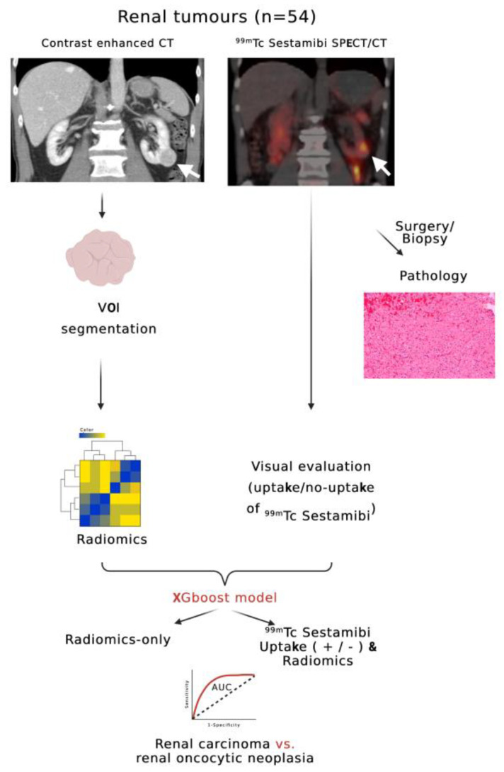
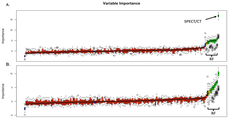
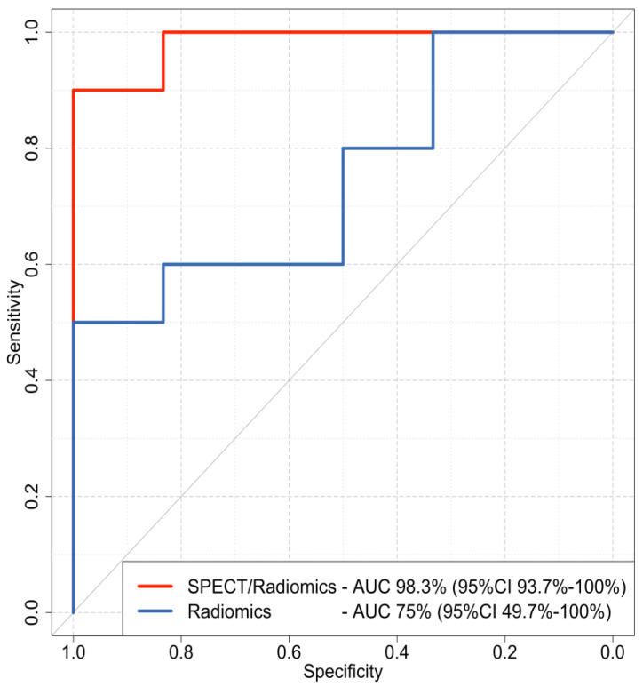
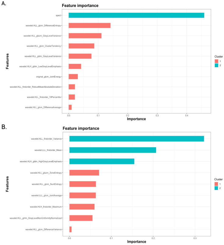
The increasing evidence of oncocytic renal tumors positive in 99mTc Sestamibi Single Photon Emission Tomography/Computed Tomography (SPECT/CT) examination calls for the development of diagnostic tools to differentiate these tumors from more aggressive forms. This study combined radiomics analysis with the uptake of 99mTc Sestamibi on SPECT/CT to differentiate benign renal oncocytic neoplasms from renal cell carcinoma. A total of 57 renal tumors were prospectively collected. Histopathological analysis and radiomics data extraction were performed. XGBoost classifiers were trained using the radiomics features alone and combined with the results from the visual evaluation of 99mTc Sestamibi SPECT/CT examination. The combined SPECT/radiomics model achieved higher accuracy (95%) with an area under the curve (AUC) of 98.3% (95% CI 93.7-100%) than the radiomics-only model (71.67%) with an AUC of 75% (95% CI 49.7-100%) and visual evaluation of 99mTc Sestamibi SPECT/CT alone (90.8%) with an AUC of 90.8% (95%CI 82.5-99.1%). The positive predictive values of SPECT/radiomics, radiomics-only, and 99mTc Sestamibi SPECT/CT-only models were 100%, 85.71%, and 85%, respectively, whereas the negative predictive values were 85.71%, 55.56%, and 94.6%, respectively. Feature importance analysis revealed that 99mTc Sestamibi uptake was the most influential attribute in the combined model. This study highlights the potential of combining radiomics analysis with 99mTc Sestamibi SPECT/CT to improve the preoperative characterization of benign renal oncocytic neoplasms. The proposed SPECT/radiomics classifier outperformed the visual evaluation of 99mTc Sestamibii SPECT/CT and the radiomics-only model, demonstrating that the integration of 99mTc Sestamibi SPECT/CT and radiomics data provides improved diagnostic performance, with minimal false positive and false negative results.
Keywords: 99mTc Sestamibi SPECT/CT; XGboost; artificial intelligence; machine learning; radiomics; renal cell carcinoma; renal oncocytic neoplasia; renal oncocytoma.
Conflict of interest statement
The authors declare no conflict of interest.
Figures
Similar articles
-
99mTc-Sestamibi SPECT/CT and histopathological features of oncocytic renal neoplasia.Scand J Urol. 2022 Oct-Dec;56(5-6):375-382. doi: 10.1080/21681805.2022.2119273. Epub 2022 Sep 5. Scand J Urol. 2022. PMID: 36065481
-
Prospective Evaluation of (99m)Tc-sestamibi SPECT/CT for the Diagnosis of Renal Oncocytomas and Hybrid Oncocytic/Chromophobe Tumors.Eur Urol. 2016 Mar;69(3):413-6. doi: 10.1016/j.eururo.2015.08.056. Epub 2015 Sep 18. Eur Urol. 2016. PMID: 26386607
-
Diagnostic Accuracy of 99mTc-Sestamibi SPECT/CT for Characterization of Solid Renal Masses.J Nucl Med. 2023 Jan;64(1):90-95. doi: 10.2967/jnumed.122.264329. Epub 2022 Jun 30. J Nucl Med. 2023. PMID: 35772963
-
Characterizing Indeterminate Renal Masses with Molecular Imaging: the Role of 99mTc-MIBI SPECT/CT.Curr Urol Rep. 2017 Sep 12;18(11):86. doi: 10.1007/s11934-017-0737-0. Curr Urol Rep. 2017. PMID: 28900880 Review.
-
Diagnostic accuracy of 99mTc-sestamibi SPECT/CT for detecting renal oncocytomas and other benign renal lesions: a systematic review and meta-analysis.Abdom Radiol (NY). 2020 Aug;45(8):2532-2541. doi: 10.1007/s00261-020-02469-8. Abdom Radiol (NY). 2020. PMID: 32193593
Cited by
-
Editorial for Special Issue "Image Analysis and Machine Learning in Cancers".Cancers (Basel). 2025 Feb 25;17(5):778. doi: 10.3390/cancers17050778. Cancers (Basel). 2025. PMID: 40075626 Free PMC article.
-
MRI-based radiomics machine learning model to differentiate non-clear cell renal cell carcinoma from benign renal tumors.Eur J Radiol Open. 2024 Oct 29;13:100608. doi: 10.1016/j.ejro.2024.100608. eCollection 2024 Dec. Eur J Radiol Open. 2024. PMID: 39525508 Free PMC article.
-
Convolutional neural networks for the differentiation between benign and malignant renal tumors with a multicenter international computed tomography dataset.Insights Imaging. 2024 Jan 25;15(1):26. doi: 10.1186/s13244-023-01601-8. Insights Imaging. 2024. PMID: 38270726 Free PMC article.
-
Small Renal Masses: Developing a Robust Radiomic Signature.Cancers (Basel). 2023 Sep 14;15(18):4565. doi: 10.3390/cancers15184565. Cancers (Basel). 2023. PMID: 37760532 Free PMC article.
References
-
- Tzortzakakis A., Papathomas T., Gustafsson O., Trpkov K., Ekström-ehn L., Arvanitis A., Karlsson M., Kokaraki G., Axelsson R., Tzortzakakis A., et al. 99mTc-Sestamibi SPECT/CT and histopathological features of oncocytic renal neoplasia. Scand. J. Urol. 2022;56:375–382. doi: 10.1080/21681805.2022.2119273. - DOI - PubMed
-
- Viswambaram P., Swarbrick N., Picardo A., Hohnen A., Pham K., Macdonald W., Hayne D., Hamid A. Technetium-99 m-sestamibi single-photon emission computerised tomography (CT)/CT in the prediction of malignant versus benign small renal masses. BJU Int. 2022;130:23–31. doi: 10.1111/bju.15737. - DOI - PubMed
-
- Warren H., Wagner T., Gorin M.A., Rowe S., Holman B.F., Pencharz D., El-Sheikh S., Barod R., Patki P., Mumtaz F., et al. Protocol for a MULTI-centre feasibility study to assess the use of 99mTc-sestaMIBI SPECT/CT in the diagnosis of kidney tumours (MULTI-MIBI study) BMJ Open. 2023;13:e067496. doi: 10.1136/bmjopen-2022-067496. - DOI - PMC - PubMed
-
- Gill A.J., Moch H., Amin M.B., Berney D.M., Compe E.M., Hartmann A., Menon S., Raspollini M.R., Rubin M.A., Srigley J.R., et al. The 2022 World Health Organization Classification of Tumours of the Urinary System and Male Genital Organs—Part A: Renal, Penile, and Testicular Tumours. Cancers. 2022;15:2155. doi: 10.1016/j.eururo.2022.06.016. - DOI - PubMed
Grants and funding
LinkOut - more resources
Full Text Sources
