. 2023 Jul 20;15(14):3689.
doi: 10.3390/cancers15143689.

Loss of PARP7 Increases Type I Interferon Signaling in EO771 Breast Cancer Cells and Prevents Mammary Tumor Growth by Increasing Antitumor Immunity

Affiliations

Loss of PARP7 Increases Type I Interferon Signaling in EO771 Breast Cancer Cells and Prevents Mammary Tumor Growth by Increasing Antitumor Immunity

Marit Rasmussen et al. Cancers (Basel). .

Abstract

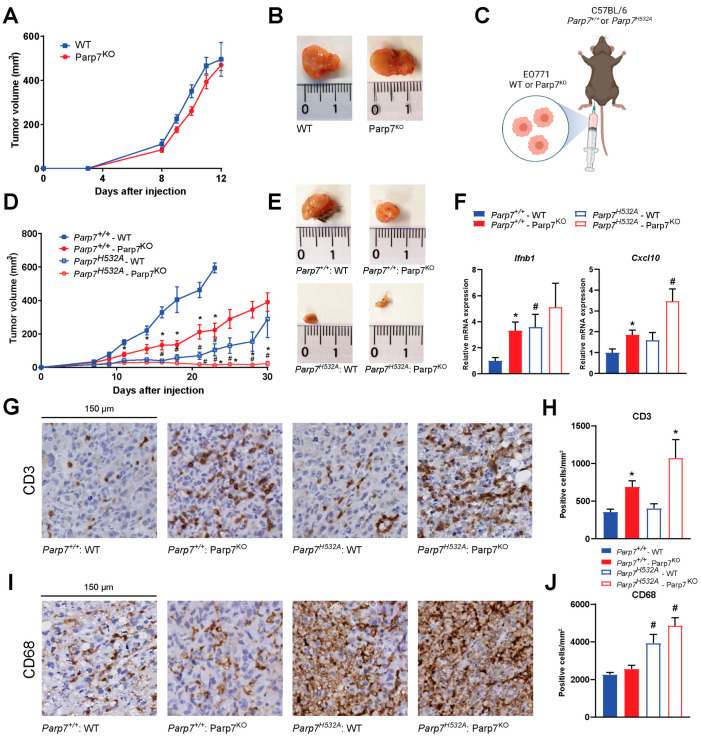
PARP7 is a member of the ADP-ribosyltransferase diphtheria toxin-like (ARTD) family and acts as a repressor of type I interferon (IFN) signaling. PARP7 inhibition causes tumor regression by enhancing antitumor immunity, which is dependent on the stimulator of interferon genes (STING) pathway, TANK-binding kinase 1 (TBK1) activity, and cytotoxic CD8+ T cells. To better understand PARP7's role in cancer, we generated and characterized PARP7 knockout (Parp7KO) EO771 mouse mammary cancer cells in vitro and in a preclinical syngeneic tumor model using catalytic mutant Parp7H532A mice. Loss of PARP7 expression or inhibition of its activity increased type I IFN signaling, as well as the levels of interferon-stimulated gene factor 3 (ISGF3) and specifically unphosphorylated-ISGF3 regulated target genes. This was partly because PARP7's modification of the RelA subunit of nuclear factor κ-B (NF-κB). PARP7 loss had no effect on tumor growth in immunodeficient mice. In contrast, injection of wildtype cells into Parp7H532A mice resulted in smaller tumors compared with cells injected into Parp7+/+ mice. Parp7H532A mice injected with Parp7KO cells failed to develop tumors and those that developed regressed. Our data highlight the importance of PARP7 in the immune cells and further support targeting PARP7 for anticancer therapy.

Keywords: ADP-ribosylation; PARP7; breast cancer; tumor immunity; type I interferon.


Conflict of interest statement

J.M. is a consultant for Duke Street Bio Inc.

Figures

Figure 1
Figure 1
EO771 cells express PARP7 and are responsive to DMXAA stimulated increases in type I interferon (IFN) signaling. (A) EO771 or (B) NCI-H1373 cells were treated with RBN-2397 for 24 h to inhibit PARP7 activity and enable visualization with Western blotting. Levels of AHR were also determined. (C) AHR levels in WT and AhrKO PyMT cells and EO771 cells were determined with Western blotting. (D) RBN-2397 treatment slightly but significantly reduces EO771 cell proliferation, whereas (E) NCI-H1373 cells are sensitive to the antiproliferative effects of RBN-2397. Cell proliferation was analyzed in an IncuCyte instrument. (F,G) EO771 cells are responsive to stimulation with the STING activator, DMXAA. Cells were treated with DMXAA for 1, 2, 4, 6, and 24 h, and relative (F) Ifnb1 and (G) Cxcl10 mRNA levels were analyzed by RT-qPCR. * Denotes statistical significance (p < 0.05) from the DMSO treated samples (D,E) or untreated samples (0 h) (F,G). Original western blots has been presented in File S1.
Figure 2
Figure 2
PARP7 loss or its inhibition increases type I IFN signaling. (A) PARP7 protein levels were determined in EO771 WT cells and Parp7KO clone 1 and 2 by Western blotting. Only the WT displays a PARP7 band in response to PARP7 inhibition. (B) Both Parp7KO clones proliferate significantly slower than the WT cells. (C) EO771 Parp7KO clones 1 and 2 form significantly smaller spheroids than the WT cells. (D) Ifnb1 mRNA levels increased in EO771 Parp7KO and RBN-2397-treated WT cells. Cells were treated with 10 µg/mL of DMXAA (+/− 100 nM RBN-2397) for 2 h, and relative mRNA levels were measured with RT-qPCR. (E) IFN-β protein levels are higher after 4 h treatment of WT cells with DMXAA +/− RBN-2397 and DMXAA in EO771 Parp7KO cells. (F) Cxcl10 mRNA levels increased after 6 h DMXAA treatment in WT and Parp7KO cells, with higher levels in WT cells co-treated with DMXAA and RBN-2397 compared with DMXAA-treated Parp7KO cells. (G) Ifnb1 levels increased in both untreated and DMXAA-treated Parp7H532A MEFs, and in WT MEFs co-treated with DMXAA and RBN-2397. (H) Cxcl10 mRNA levels increased after 6 h treatment with DMXAA Parp7H532A and WT MEFs, with higher levels in WT MEFs co-treated with DMXAA + RBN-2397 compared with DMXAA Parp7H532A MEFs. (I) Parp7KO cells display elevated constitutive Pdl1 levels compared with WT cells treated +/− DMXAA. (J) DMXAA treatment results in a significant decrease in proliferation in the EO771 WT and Parp7KO cells, with greater effect observed in Parp7KO cells. (K) Heatmap showing expression levels of different PARPs in response to DMXAA and/or RBN-2397 in EO771 WT and Parp7KO cells. Cells were treated with 10 µg/mL of DMXAA (+/− 100 nM RBN-2397) for 2 h, and relative mRNA levels were measured with RT-qPCR. * Denotes statistical significance (p < 0.05) compared to DMSO; # (for (B) these are colored to indicate which graph they correspond to) denotes statistical significance due to loss the of PARP7 activity. Original western blots has been presented in File S1.
Figure 3
Figure 3
Levels of unphosphorylated interferon stimulated gene factor 3 (ISGF3) are upregulated in EO771 Parp7KO cells. (A) EO771 WT and Parp7KO cells have intact STING regulated type I IFN signaling pathway. The expression and/or phosphorylation of cGAS, STING, TBK1 and IRF3 are similar between WT and Parp7KO cells. However, Parp7KO cells have increased levels of unphosphorylated signal transducer and activator of transcription 1 (STAT1), STAT2, and interferon regulatory factor 9 (IRF9). Cells were treated with 10 µg/mL of DMXAA (+/− 100 nM RBN-2397) for 0, 2, 6, or 24 h, and samples were subjected to SDS-PAGE and Western blotting. (B) Levels of native STAT1 are higher in the EO771 Parp7KO cells independent of DMXAA treatment. (C) Levels of phosphorylated STAT1 after DMXAA treatment relative to native STAT1 are lower in the EO771 Parp7KO cells compared with both WT and RBN-2397-treated WT cells. (D) EO771 Parp7KO cells have increased cytoplasmic and nuclear levels of STAT1, STAT2, and IRF9 in both untreated and treated samples. (E) Stat1, (F) Stat2, and (G) Irf9 mRNA levels are higher in EO771 Parp7KO cells and after 24 h RBN-2397 treatment in WT cells. (H) Expression levels of P-ISGF3 target genes Myd88 is not significantly affected by PARP7 loss, while target genes (I) Adar and (J) Irf1 are slightly higher in the EO771 Parp7KO cells. (G) Expression levels of U-ISGF3 target genes, (K) Isg15 and (L) Mx1, and (M) ISG15-induced gene Usp18 are all significantly higher in EO771 Parp7KO cells and RBN-2397-treated WT cells. Cells were treated with 10 µg/mL of DMXAA (+/− 100 nM RBN-2397) for 6 h, and mRNA levels were quantified with RT-qPCR. * Denotes statistical significance (p < 0.05) compared to no treatment (EG) or DMSO (HM); # denotes statistical significance due to the loss of PARP7. Original western blots have been presented in File S1.
Figure 4
Figure 4
Nuclear factor κB (NF-κB) is involved in the increased type I IFN signaling observed in EO771 Parp7KO cells. (A) Inhibition of TANK-binding kinase 1 (TBK1) with MRT67307 significantly decreases Ifnb1 mRNA levels in EO771 Parp7KO cells but not in WT cells. Inhibition of both TBK1 and the IκB kinases (IKKs) with BX795 blocks Ifnb1 mRNA expression in all cases. Cells were treated with either 1 µM of MRT67307 or 100 nM of BX795 for 24 h, followed by 10 µg/mL of DMXAA for 2 h. (B) Treatment with DMXAA and/or RBN-2397 increases protein levels of both p50 and RelA in EO771 WT cells while levels are higher in Parp7KO cells in both untreated and treated cells. Cells were treated with 10 µg/mL of DMXAA (+/− 100 nM of RBN-2397) for 24 h and proteins were visualized by Western blotting. (C) Levels of expressed NF-κB subunits are higher in response to PARP7 inhibition, and levels of RelA are significantly higher in the Parp7KO cells. Protein levels of p50 and RelA in untreated samples were quantified by normalizing to the loading control. (D) Expression levels of Rela mRNA are not affected by DMXAA or PARP7. (E) Knockdown of either TBK1, RelA, or both, in EO771 WT and Parp7KO cells. After 48 h of transfection with 25 nM of siRNA targeting Tbk1, Rela, or both, protein levels were visualized by Western blotting. (F) Knockdown of TBK1 and RelA results in significantly decreased Ifnb1 mRNA levels in Parp7KO cells but not in WT cells. After 48 h of transfection with 25 nM of siRNA for Tbk1, Rela, or both, cells were treated with 10 µg/mL of DMXAA for 2 h. (G) p50 and RelA interact with WT and catalytically inactive PARP7, and RelA is MARylated by WT PARP7. Cos-1 cells were transfected with FLAG-tagged p50 or RelA together with either GFP-tagged WT PARP7 or the catalytically inactive H532A mutant. The FLAG-tagged p50 or RelA was immunoprecipitated, and the interaction and modification was examined by Western blotting. * Denotes statistical significance (p < 0.05) compared to DMSO; # denotes statistical difference due to the loss of PARP7; “a” denotes statistical difference compared to no inhibitor (A) or to the nontargeting siRNA (F). Original western blots have been presented in File S1.
Figure 5
Figure 5
Loss of PARP7 prevents mammary tumor growth in immunocompetent mice. (A) No difference in tumor growth is observed between WT and Parp7KO cells injected into immunodeficient NSG-mice, n = 8. (B) Representative images of tumors dissected from NSG mice injected with WT (left) or Parp7KO (right) cells. (C) Graphical representation of experimental setup indicating injection of either WT or Parp7KO cells into either C57BL/6 Parp7+/+ or Parp7H532A mice. Figure made with BioRender. (D) Loss of PARP7 in the cancer cells significantly decreases tumor growth in Parp7+/+ mice, and this is further decreased in Parp7H532A mice, n = 6–12. * Denotes statistical significance (p < 0.05) between the genotype of the cells injected, while # denotes statistical significance between the genotype of the mice. (E) Representative images of tumors dissected from Parp7+/+ mice injected with WT or Parp7KO cells and from Parp7H532A mice injected with WT or Parp7KO cells, respectively. (F) Tumor expression levels of Ifnb1 and Cxcl10 are increased in response to loss of PARP7 activity in either cancer cells or in recipient immune system. * Denotes statistical significance (p < 0.05) compared to the WT cells injected in the corresponding mouse strain, and # denotes statistical significance compared to corresponding cell line injected in the Parp7+/+ mice. (G) Injection of Parp7KO cells increases tumor infiltration of T cells. Representative images of tumor sections stained with antibody against CD3. (H) Quantification of T-cell infiltration with CD3-positive cells per mm2, n = 8. (I) Loss of functional PARP7 in recipient mice results in increased tumor infiltration of macrophages. Representative images of tumor sections stained with antibody against CD68. (J) Quantification of macrophage infiltration, n = 8. For (H,J), # denotes statistical significance (p < 0.05) between the genotype of the mice injected, while * denotes significance between the cells injected.
Figure 6
Figure 6
Loss of PARP7 increases type I IFN signaling in CD8+ T cells and increases M1 macrophage signaling. (A) Enrichment of Cd8a mRNA verifies CD8+ T-cell isolation. Levels of Cd8a mRNA expression is increased in isolated CD8+ T cells compared to the splenocyte starting material. Expression levels were measured with RT-qPCR. (B) Levels of type I IFN Ifnb1 are significantly increased in both resting and activated CD8+ T cells isolated from Parp7H532A mice. Cells were either kept in a resting state or activated with CD3/CD28 beads prior to treatment with 10 µg/mL of DMXAA for 24 h. (C) Loss of PARP7 function results in higher secreted levels of IFN-β from activated CD8+ T cells treated with 10 µg/mL of DMXAA for 24 h. (D) The levels of the type II IFN Ifng do not significantly differ between the genotypes, while (E) the levels of the type III IFN Ifnl2 are significantly upregulated in activated CD8+ T cells isolated from Parp7H532A mice. (F) Levels of Il6 and (G) Ifnb1 are higher in M1 macrophages isolated from Parp7H532A mice compared with Parp7+/+ mice. (H) M2 macrophages from Parp7H532A mice have higher expression levels of Arg1. For (FH), bone-marrow-derived macrophages were isolated and polarized, and the mRNA levels were determined with RT-qPCR and normalized to the M0. * Denotes statistical significance (p < 0.05) compared to splenocytes (A) or DMSO (BE); # denotes statistical significance due to loss of PARP7 function (B,C,EH); “a” denotes statistical significance compared to the resting cells (B,D,E) or the unpolarized M0 macrophages (FH). n = 3.
Figure 7
Figure 7
Proposed mechanism of action. (A) In the presence of cytoplasmic DNA, cGAS is activated and catalyzes the synthesis of cGAMP, which activates STING. STING subsequently activates TBK1, which phosphorylates and activates IRF3. STING activation also results in the release of NF-κB from IκBα. Both pIRF3 and NF-κB translocate to the nucleus and bind to the promoter region of type I IFNs, such as Ifnb1. PARP7 inhibits the activity of both TBK1 and the RelA subunit of NF-κB. Secreted IFN-β binds to IFNAR, which results in phosphorylation of STAT1 and STAT2. Together with IRF9, they form P-ISGF3 which regulates the expression of ISGs. Upon sustained IFN-β, unphosphorylated ISGF3 is upregulated, which results in transcription of certain ISGs. (B) Loss of PARP7 results in increased production and secretion of IFN-β, which further increases both P-ISGF3 and U-ISGF3 signaling. Figure made in BioRender (license agreement YX25IUCM4M to J.M.).

Similar articles

Cited by

References

    1. Shergold A.L., Millar R., Nibbs R.J.B. Understanding and overcoming the resistance of cancer to PD-1/PD-L1 blockade. Pharmacol. Res. 2019;145:104258. doi: 10.1016/j.phrs.2019.104258. - DOI - PubMed
    1. Li T., Chen Z.J. The cGAS-cGAMP-STING pathway connects DNA damage to inflammation, senescence, and cancer. J. Exp. Med. 2018;215:1287–1299. doi: 10.1084/jem.20180139. - DOI - PMC - PubMed
    1. Cheon H., Borden E.C., Stark G.R. Interferons and their stimulated genes in the tumor microenvironment. Semin. Oncol. 2014;41:156–173. doi: 10.1053/j.seminoncol.2014.02.002. - DOI - PMC - PubMed
    1. Parker B.S., Rautela J., Hertzog P.J. Antitumour actions of interferons: Implications for cancer therapy. Nat. Rev. Cancer. 2016;16:131–144. doi: 10.1038/nrc.2016.14. - DOI - PubMed
    1. Chen Q., Sun L., Chen Z.J. Regulation and function of the cGAS-STING pathway of cytosolic DNA sensing. Nat. Immunol. 2016;17:1142–1149. doi: 10.1038/ni.3558. - DOI - PubMed