. 2023 Jul 6;11(7):1909.
doi: 10.3390/biomedicines11071909.

GDF8 Contributes to Liver Fibrogenesis and Concomitant Skeletal Muscle Wasting

Affiliations

GDF8 Contributes to Liver Fibrogenesis and Concomitant Skeletal Muscle Wasting

Alexander Culver et al. Biomedicines. .

Abstract

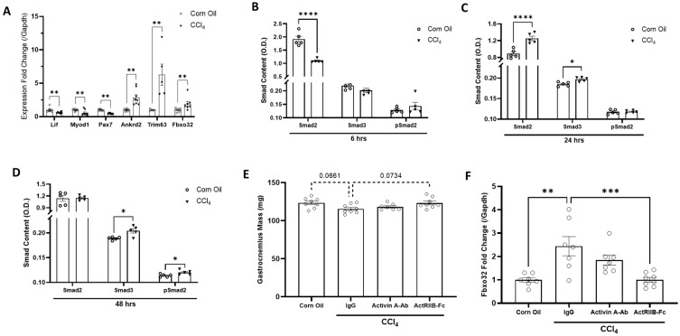
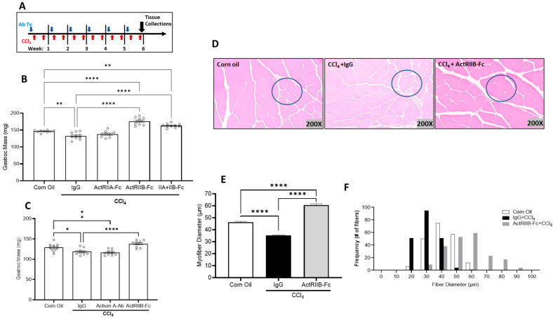
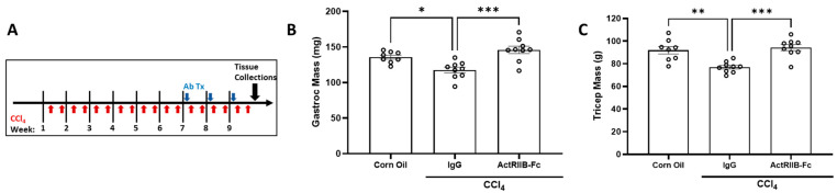
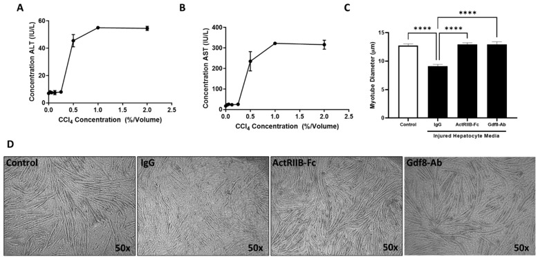
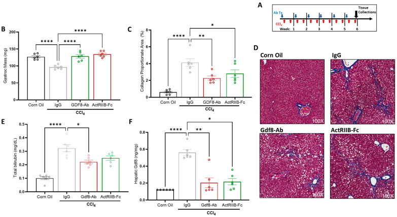
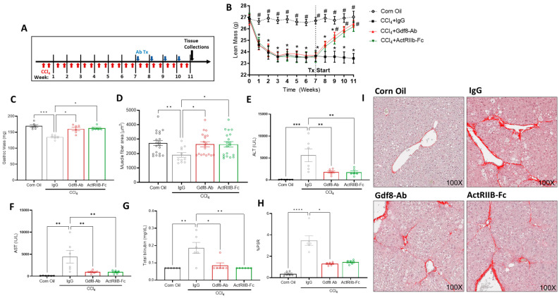
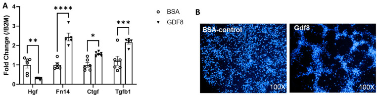
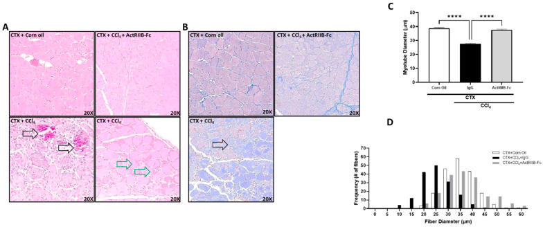
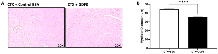
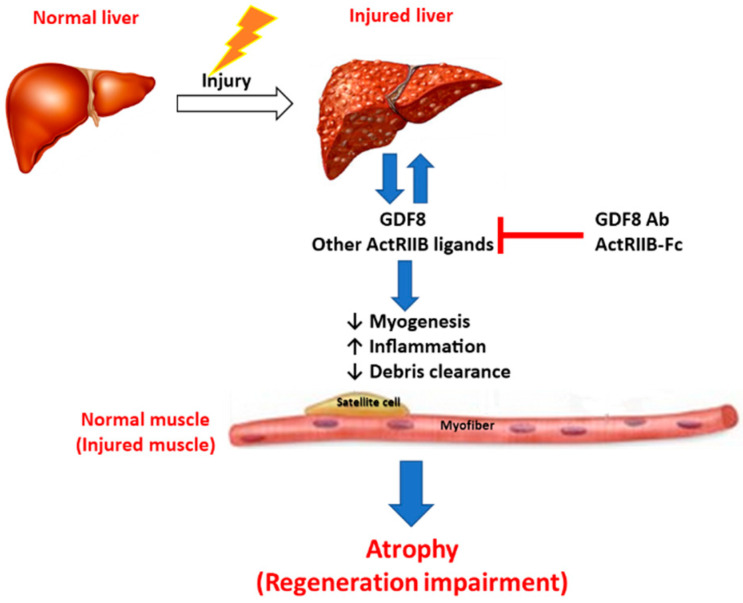
Patients with end-stage liver disease exhibit progressive skeletal muscle atrophy, highlighting a negative crosstalk between the injured liver and muscle. Our study was to determine whether TGFβ ligands function as the mediators. Acute or chronic liver injury was induced by a single or repeated administration of carbon tetrachloride. Skeletal muscle injury and repair was induced by intramuscular injection of cardiotoxin. Activin type IIB receptor (ActRIIB) ligands and growth differentiation factor 8 (Gdf8) were neutralized with ActRIIB-Fc fusion protein and a Gdf8-specific antibody, respectively. We found that acute hepatic injury induced rapid and adverse responses in muscle, which was blunted by neutralizing ActRIIB ligands. Chronic liver injury caused muscle atrophy and repair defects, which were prevented or reversed by inactivating ActRIIB ligands. Furthermore, we found that pericentral hepatocytes produce excessive Gdf8 in injured mouse liver and cirrhotic human liver. Specific inactivation of Gdf8 prevented liver injury-induced muscle atrophy, similar to neutralization of ActRIIB ligands. Inhibition of Gdf8 also reversed muscle atrophy in a treatment paradigm following chronic liver injury. Direct injection of exogenous Gdf8 protein into muscle along with acute focal muscle injury recapitulated similar dysregulated muscle regeneration as that observed with liver injury. The results indicate that injured liver negatively communicate with the muscle largely via Gdf8. Unexpectedly, inactivation of Gdf8 simultaneously ameliorated liver fibrosis in mice following chronic liver injury. In vitro, Gdf8 induced human hepatic stellate (LX-2) cells to form a septa-like structure and stimulated expression of profibrotic factors. Our findings identified Gdf8 as a novel hepatomyokine contributing to injured liver-muscle negative crosstalk along with liver injury progression.

Keywords: Gdf8; TGFβ family; liver injury; liver–muscle crosstalk; muscle atrophy.


Conflict of interest statement

The authors declare no conflict of interest.

Figures

Figure 1
Figure 1
CCl4-induced liver injury acutely activates muscle catabolic gene markers and downregulates satellite cell markers. Female mice were administered a single dose of corn oil or carbon tetrachloride (CCl4). Muscle samples were collected at 6, 24, and 48 h post CCl4 injection. (A) Muscle mRNA expression of the genes indicated was analyzed 6 h after livery injury. (BD) Protein levels of muscle total Smad 2 and Smad 3 and phosphorylated Smad 2 in muscle were measured via ELISA 6, 24, and 48 h post injury. In a subsequent study, female mice received IgG, activin A-Ab, or ActRIIB-Fc (10 mg/kg) 16 h prior to a single administration of CCl4. (E) Gastrocnemius muscle wet weight and real-time PCR analysis of (F) muscle Fbxo32 78 h following CCl4 injection are shown. Data are expressed as mean ± SEM (n = 5 mice/group). Significance is indicated as * p ≤ 0.05, ** p ≤ 0.01, *** p ≤ 0.001, or **** p ≤ 0.0001. p-values between 0.05 and 0.1 are indicated by dotted lines.
Figure 2
Figure 2
CCl4-induced chronic liver injury induces muscle atrophy in mice independent of sex. (A) Male mice were chronically administered carbon tetrachloride (CCl4) twice a week for 6 weeks. Sixteen hours before the 1st CCl4 injection each week, male mice received corn oil, immunoglobulin (IgG), ActRIIA-Fc, ActRIIB-Fc, or a combination of ActRIIA-Fc and ActRIIB-Fc (10 mg/kg). A corn oil (non-injured) group was added as a homeostasis control. After 6 weeks of CCl4 treatment, (B) gastrocnemius wet weights were evaluated. Female mice underwent the same CCl4 injury paradigm for 6 weeks while receiving IgG, Activin A-Ab, or ActRIIB-Fc weekly. (C) Gastrocnemius mass was assessed by wet weight. (D) Representative H&E cross-sectional images of myofibers in the gastrocnemius muscle. (E) Average fiber diameter and (F) the frequency distribution of gastrocnemius muscle fibers were analyzed. Data are expressed as mean ± SEM (n = 10). Significance is indicated as * p ≤ 0.05, ** p ≤ 0.01, or **** p ≤ 0.0001. All quantifications of myofibers (~200 myofibers counted per group) were determined using ImageScope 12.3 software (Aperio).
Figure 3
Figure 3
Skeletal muscle mass loss induced by existing CCl4 injury was reversed with ActRIIB inhibition. (A) Female mice were injected with CCl4 or corn oil twice per week for 6 weeks. Then, mice were dosed with ActRIIB-Fc or IgG once per week for 3 weeks, during which CCl4 or corn oil injections continued (9 weeks total). (B) Gastrocnemius and (C) triceps brachii muscle masses were assessed by wet weight at study completion. Data are expressed as mean ± SEM (n = 9). Significance is indicated as * p ≤ 0.05, ** p ≤ 0.01, or *** p ≤ 0.001.
Figure 4
Figure 4
Hepatic Gdf8 content is increased in human patients with end-stage liver diseases (ESLD) similar to acutely injured mouse liver. (A) Liver samples were collected from healthy individuals (n = 5) or patients with established cirrhosis (n = 7). Liver lysates were prepared and subjected to quantification of Gdf8 protein via ELISA. All normal control samples were below quantifiable limits of the assay. (BE) Male mice (n = 5) were administered a single dose of corn oil or carbon tetrachloride (CCl4). The concentration of Gdf8 protein was measured via ELISA in the (B) liver, (C) plasma, and (D) muscle at the time points indicated after CCl4 injection. Liver sections were generated from mice 48 h post single CCl4 injection and immune-stained for Gdf8. (E) Representative images of Gdf8 immunostaining are shown. Data are expressed as mean ± SEM. Significance is indicated as * p ≤ 0.05, ** p ≤ 0.01.
Figure 5
Figure 5
Damaged primary hepatocytes release Gdf8, which directly acts on C2C12 cells. Primary mouse hepatocytes were exposed to various concentrations of CCl4 as indicated for 24 h. (A) ALT and (B) AST in the culture medium were analyzed. Hepatocyte media were collected following exposure to 0.5% CCl4 by volume. The hepatocyte media were then diluted 1:10 into C2C12 differentiation media daily for 5 days along with IgG, ActRIIB-Fc, or Gdf8-Ab (100 ng/mL for each). (C) Myotube diameter was measured by ImageScope software (Aperio) after 5 days of myotube differentiation. (D) Representative images of differentiation myotubes. Data are expressed as mean ± SEM (n = 3 wells/group). Significance is indicated as **** p ≤ 0.0001.
Figure 6
Figure 6
Gdf8-Ab or ActRIIB-Fc prevents muscle atrophy and hepatic fibrogenesis in CCl4-induced chronic liver injury. (A) Male C57BL/6 mice were chronically administered carbon tetrachloride (CCl4) twice a week for 6 weeks. Sixteen hours before the first CCl4 injection each week mice received IgG, Gdf8-Ab, or ActRIIB-Fc. A corn oil (non-injured) group was added as a homeostasis control. After 6 weeks of CCl4 injury, (B) gastrocnemius mass, (C,D) hepatic collagen proportionate area, and (E) total bilirubin levels were quantified and (F) hepatic Gdf8 levels were measured via ELISA. The minimally detectable values were used for the corn oil control group. (D) Representative images of Masson’s trichrome staining on liver sections. Data are expressed as mean ± SEM (n = 6). Significance is indicated as * p ≤ 0.05, ** p ≤ 0.01, or **** p ≤ 0.0001. Collagen proportionate area was quantified using HALO image analysis software.
Figure 7
Figure 7
Gdf8-Ab or ActRIIB-Fc reverses muscle atrophy and hepatic fibrogenesis in CCl4-induced chronic liver injury. (A) Male C57Bl/6 mice were chronically administered CCl4 or corn oil twice a week for 11 weeks. Starting in week 7, 16 h before the first weekly CCl4 injection, mice received a weekly dose of IgG, Gdf8-Ab, or ActRIIB-Fc with (B) continual total body lean mass quantification via QNMR. After 4 weeks of CCl4 + antibody treatment, (C) gastrocnemius mass and (D) muscle fiber cross-sectional areas were assessed. Plasma (E) ALT, (F) AST, and (G) total bilirubin were measured in terminal blood. (H) Percent area of collagen stain was analyzed from images of Picrosirius red staining on liver sections. (I) Representative Picrosirius red staining on liver sections are shown. Data are expressed as mean ± SEM (n = 10). Significance is indicated as * p ≤ 0.05 compared to corn oil control and as # p ≤ 0.05 vs. IgG for LBM QNMR data. For all other figures, significance is indicated as * p ≤ 0.05, ** p ≤ 0.01, *** p ≤ 0.001, or **** p ≤ 0.0001. The collagen proportionate area was quantified using HALO image analysis software.
Figure 8
Figure 8
Gdf8 activates human hepatic (LX-2) cells in vitro. Human hepatic stellate cells (LX-2) were treated with bovine serum albumin (BSA) or Gdf8 protein at equal concentrations (100 ng/mL) for 24 h. (A) Real-time PCR analyses of Hgf, Fn14, Ctgf, and Tgfb1 mRNA expression were performed. Data are expressed as mean ± SEM (n = 6). Significance is indicated as * p ≤ 0.05, ** p ≤ 0.01, *** p ≤ 0.001, or **** p ≤ 0.0001. (B) Representative DAPI-stained images (100X) of LX-2 cells. B2M, β2-microglobulin.
Figure 9
Figure 9
CCl4-induced liver injury negatively affects muscle repair following cardiotoxin (CTX) injury. Female mice were injected with carbon tetrachloride (CCl4) or corn oil every three days over a span of 10 days (3 administrations). Focal muscle injury was induced via direct CTX injection in the gastrocnemius muscle 6 h after receiving the first CCl4 treatment. ActRIIB-Fc or IgG was dosed prior to CTX injection. Muscle samples were harvested at day 10 post CTX injection. (A) Representative H&E cross-sectional images of myofibers in the gastrocnemius muscle. Black arrows indicate areas of calcification (bottom left) and green arrows indicate defective myocyte regeneration (bottom right). (B) Representative trichrome staining of muscle sections (20X). Arrow indicates fibrotic regions. (C) Fiber diameter of gastrocnemius muscles and (D) the frequency distribution of corresponding fibers (white—corn oil control, black—CCl4, and light gray—ActRIIB-Fc) were evaluated. All quantifications of myofibers (~200 counted per group) were determined using ImageScope 12.3 software (Aperio). Data are expressed as mean ± SEM (n = 6). Significance is indicated as **** p ≤ 0.0001.
Figure 10
Figure 10
Exogenous Gdf8 disrupts muscle regeneration. In female mice, muscle injury was induced by CTX, and 2 h later, 5 µg of Gdf8 or bovine serum albumin (BSA) were directly injected into the muscle. The next day, 1 µg of each was injected to mimic the injury-mediated waning in ligand exposure. Muscle samples were collected at day 10 post CTX injury. (A) Representative H&E cross-sectional images of myofibers in the gastrocnemius muscle. (B) Fiber diameter of gastrocnemius muscles. All quantifications of myofibers (~200 counted per group) were determined using ImageScope 12.3 software (Aperio). Data are expressed as mean ± SEM (n = 5). Significance is indicated as **** p ≤ 0.0001.
Figure 11
Figure 11
Hypothesis of liver and skeletal muscle communication under pathological conditions. Injured liver produces and releases Gdf8 and potentially other ActRIIB ligands, promoting liver injury progression and simultaneously causing systemic disruption of TGFβ signaling in skeletal muscle and likely other organs. ActRIIB ligands, mainly Gdf8, induce myofiber atrophy and inhibit myogenesis, as well as various biological processes, including inflammation and tissue remodeling, resulting in muscle degeneration and regeneration impairment. Degenerating skeletal muscle may in turn negatively feedback onto both the liver and skeletal muscle, promoting the progression of liver injury/fibrosis and concomitant muscle atrophy. Thus, simultaneous inhibition of ActRIIB ligands, especially Gdf8, is a powerful approach to preventing or reversing muscle atrophy concomitant to liver injury and even improving both injured liver and degenerating muscle.

Similar articles

Cited by

References

    1. Kruszynska Y., Williams N., Perry M., Home P. The relationship between insulin sensitivity and skeletal muscle enzyme activities in hepatic cirrhosis. Hepatology. 1988;6:1615–1619. doi: 10.1002/hep.1840080624. - DOI - PubMed
    1. Hsu C.S., Kao J.H. Sarcopenia and chronic liver diseases. Expert Rev. Gastroenterol. Hepatol. 2018;12:1229–1244. doi: 10.1080/17474124.2018.1534586. - DOI - PubMed
    1. Bhanji R.A., Narayanan P., Allen A.M., Malhi H., Watt K.D. Sarcopenia in hiding: The risk and consequence of underestimating muscle dysfunction in nonalcoholic steatohepatitis. Hepatology. 2017;66:2055–2065. doi: 10.1002/hep.29420. - DOI - PubMed
    1. Koo B.K., Kim D., Joo S.K., Kim J.H., Chang M.S., Kim B.G., Lee K.L., Kim W. Sarcopenia is an independent risk factor for non-alcoholic steatohepatitis and significant fibrosis. J. Hepatol. 2017;66:123–131. doi: 10.1016/j.jhep.2016.08.019. - DOI - PubMed
    1. Lee Y.H., Kim S.U., Song K., Park J.Y., Kim D.Y., Ahn S.H., Lee B.W., Kang E.S., Cha B.S., Han K.H. Sarcopenia is associated with significant liver fibrosis independently of obesity and insulin resistance in nonalcoholic fatty liver disease: Nationwide surveys (KNHANES 2008–2011) Hepatology. 2016;63:776–786. doi: 10.1002/hep.28376. - DOI - PubMed

LinkOut - more resources