Review
. 2023 Jul 6;11(7):1922.
doi: 10.3390/biomedicines11071922.

Angiogenesis and Tissue Repair Depend on Platelet Dosing and Bioformulation Strategies Following Orthobiological Platelet-Rich Plasma Procedures: A Narrative Review

Affiliations
Review

Angiogenesis and Tissue Repair Depend on Platelet Dosing and Bioformulation Strategies Following Orthobiological Platelet-Rich Plasma Procedures: A Narrative Review

Peter A Everts et al. Biomedicines. .

Abstract

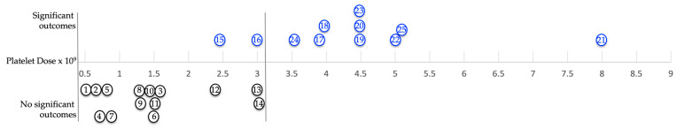
Angiogenesis is the formation of new blood vessel from existing vessels and is a critical first step in tissue repair following chronic disturbances in healing and degenerative tissues. Chronic pathoanatomic tissues are characterized by a high number of inflammatory cells; an overexpression of inflammatory mediators; such as tumor necrosis factor-α (TNF-α) and interleukin-1 (IL-1); the presence of mast cells, T cells, reactive oxygen species, and matrix metalloproteinases; and a decreased angiogenic capacity. Multiple studies have demonstrated that autologous orthobiological cellular preparations (e.g., platelet-rich plasma (PRP)) improve tissue repair and regenerate tissues. There are many PRP devices on the market. Unfortunately, they differ greatly in platelet numbers, cellular composition, and bioformulation. PRP is a platelet concentrate consisting of a high concentration of platelets, with or without certain leukocytes, platelet-derived growth factors (PGFs), cytokines, molecules, and signaling cells. Several PRP products have immunomodulatory capacities that can influence resident cells in a diseased microenvironment, inducing tissue repair or regeneration. Generally, PRP is a blood-derived product, regardless of its platelet number and bioformulation, and the literature indicates both positive and negative patient treatment outcomes. Strangely, the literature does not designate specific PRP preparation qualifications that can potentially contribute to tissue repair. Moreover, the literature scarcely addresses the impact of platelets and leukocytes in PRP on (neo)angiogenesis, other than a general one-size-fits-all statement that "PRP has angiogenic capabilities". Here, we review the cellular composition of all PRP constituents, including leukocytes, and describe the importance of platelet dosing and bioformulation strategies in orthobiological applications to initiate angiogenic pathways that re-establish microvasculature networks, facilitating the supply of oxygen and nutrients to impaired tissues.

Keywords: angiogenesis; bioformulation; biosurgery; leukocytes; orthobiology; platelet dose; platelet-rich fibrin; platelet-rich plasma; tissue repair.


Conflict of interest statement

Peter A. Everts is also the Chief Scientific Officer for EmCyte Corporation.

Figures

Figure 2
Figure 2
Whole blood cellular gravitational density separation after a dual-spin procedure. In this picture, a 60 mL PRP device (PurePRP-SP®, EmCyte Corporation, Fort Myers FL, USA, with permission) was used to produce LR-PRP. At A, a thin, gray, multicomponent, buffy-coat layer is visible. Before extraction of this layer, platelets are resuspended in a small volume of plasma and aspirated in a syringe. Resuspending the platelets colors the PRP slightly light amber, indicating a capture of the buffy-coat layer. A magnified image of the buffy-coat layer in B illustrates how blood cells are arranged according to their densities [70]. To realize an LR-PRP specimen, a fraction of RBCs must be part of the bioformulation, as the density of neutrophils overlaps with the density of the RBCs. In this 3.75 mL LR-PRP product, the total available platelets were 10.02 × 10⁹; monocyte and neutrophil concentrations were 7.95 and 9.92 × 10³/µL, respectively. The RBC concentration was 2.01 × 10⁶/µL.
Figure 1
Figure 1
PRP-G clot. A liquid, 7 mL PRP concentrate specimen, was mixed with a combination of 0.30 mL of CaCl (10%) and 0.2 mL of thrombin (50 IU) to induce the polymerization of fibrinogen, which is present in the PRP plasma fraction, into fibrin. The result is a stable PRP-G scaffold that contains high concentrations of platelets and eventually leukocytic cells, when compared to PRF preparations [35].
Figure 3
Figure 3
An electron microscopic image of an ultrathin (70 nm sample) PRP fraction. In the red circles, individual non-activated platelets are visible. In the blue circles, platelets have released their content following platelet activation. A single, non-activated, platelet at magnification ×7000 visualizes the three different platelet granules. Abbreviations: α: alpha granule; DG: dense granule; L: lysosome.
Figure 4
Figure 4
An illustration of an activated platelet, indicating pro- and anti-angiogenic platelet constituents released from α and dense granules. Abbreviations: TGF-β1: transforming growth factor beta 1; VEGF-A: vascular endothelial growth factor-ligand A; SDF-1α: stromal cell-derived factor-1 alpha; Ang-1: angiopoietin-1; IL-8: interleukin-8; MMP: matrix metalloproteases; PMP: platelet microparticles; TNF-α: tumor necrosis factor-alpha; 5-HT: serotonin; PDGF-BB: platelet-derived growth factor-BB; bFGF: basic fibroblast growth factor; TSP-1: thrombospondin 1; TIMP: tissue inhibitor of metalloproteinase; PF4: platelet factor 4; RANTES: regulated by T-cell activation and probably secreted by T cells.
Figure 5
Figure 5
Platelet- and leukocyte-mediated angiogenic pathways.
Figure 6
Figure 6
The effectiveness of PRP products was studied for a variety of orthobiological soft tissue pathoanatomic conditions, including tendinopathies, hamstring pathologies, meniscus lesions, and plantar fasciitis. Each of these studies, represented by the circles, has the platelet dose depicted on the X-axis (e.g., in study 24, the platelet dose was on average 3.6 × 10⁹ platelets for all treated patients). Based on the assessment scores between the treatment groups in each study, the Y-axis indicates whether the patient outcomes were positive or negative. It was found that results of studies with identifiers 1–14 were not significantly different when compared with control groups, particularly when platelet doses were less than 1.5 × 10⁹ platelets in the majority of these studies. All markers in blue demonstrated a positive outcome after PRP treatment. A notable characteristic of these studies is that the platelet dose was generally higher than that observed in studies without significant outcomes. Studies involving greater than 3.2 × 10⁹ platelets have generally reported more positive results. (Based on the data provided in each study, the platelet dose was calculated; however, if no platelet count was provided, the baseline count was assumed to be 250.000/µL for all studies. The platelet capture rates and concentration factors were determined using the methods described by Magalon et al. and Fadadu et al. [2,4]).

References

    1. Everts P.A., Flanagan G., Podesta L. Autologous Orthobiologics. In: Mostoufi S.A., George T.K., Tria A.J. Jr., editors. Clinical Guide to Musculoskeletal Medicine: A Multidisciplinary Approach. Springer International Publishing; Cham, Switzerland: 2022. pp. 651–679.
    1. Fadadu P.P., Mazzola A.J., Hunter C.W., Davis T.T. Review of Concentration Yields in Commercially Available Platelet-Rich Plasma (PRP) Systems: A Call for PRP Standardization. Reg. Anesth. Pain Med. 2019;44:652–659. doi: 10.1136/rapm-2018-100356. - DOI - PubMed
    1. Everts P., Onishi K., Jayaram P., Lana J.F., Mautner K. Platelet-Rich Plasma: New Performance Understandings and Therapeutic Considerations in 2020. Int. J. Mol. Sci. 2020;21:7794. doi: 10.3390/ijms21207794. - DOI - PMC - PubMed
    1. Magalon J., Brandin T., Francois P., Degioanni C., De Maria L., Grimaud F., Veran J., Dignat-George F., Sabatier F. Technical and Biological Review of Authorized Medical Devices for Platelets-Rich Plasma Preparation in the Field of Regenerative Medicine. Platelets. 2021;32:200–208. doi: 10.1080/09537104.2020.1832653. - DOI - PubMed
    1. Gentile P., Garcovich S. Systematic Review—The Potential Implications of Different Platelet-Rich Plasma (PRP) Concentrations in Regenerative Medicine for Tissue Repair. Int. J. Mol. Sci. 2020;21:5702. doi: 10.3390/ijms21165702. - DOI - PMC - PubMed