

[18F]FDG-PET/CT in Idiopathic Inflammatory Myopathies: Retrospective Data from a Belgian Cohort
- PMID: 37510060
- PMCID: PMC10377909
- DOI: 10.3390/diagnostics13142316
[18F]FDG-PET/CT in Idiopathic Inflammatory Myopathies: Retrospective Data from a Belgian Cohort
Abstract
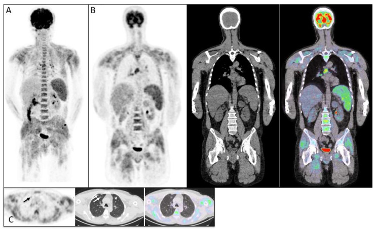
[18F]FDG-PET/CT is a useful tool for diagnosis and cancer detection in idiopathic inflammatory myopathies (IIMs), especially polymyositis (PM) and dermatomyositis (DM). Data deriving from Europe are lacking. We describe [18F]FDG-PET/CT results in a Belgian cohort with IIMs, focusing on patients with PM and DM. All of the cases of IIMs admitted between December 2010 and January 2023 to the Cliniques Universitaires Saint-Luc (Belgium) were retrospectively reviewed. In total, 44 patients were identified with suspected IIMs; among them, 29 were retained for final analysis. The mean age of the retained patients was 48.7 years; 19 patients were female (65.5%). Twenty-two patients had DM and seven had PM. The mean serum creatinine kinase (CK) and the mean CRP levels were 3125 UI/L and 30.3 mg/L, respectively. [18F]FDG-PET/CT imaging was performed for 27 patients, detecting interstitial lung diseases (ILDs) in 7 patients (25.9%), cancer in 3 patients (11.1%), and abnormal muscle FDG uptake compatible with myositis in 13 patients (48.1%). All of the patients who were detected to have ILDs via PET/CT imaging were confirmed using a low-dose lung CT scan. Among the patients who were detected to have abnormal muscle FDG uptake via PET/CT scans (13/28), the EMG was positive in 12 patients (p = 0.004), while the MRI was positive in 8 patients (p = 0.02). We further observed that there was a significantly higher level of CK in the group with abnormal muscle FDG uptake (p = 0.008). Our study showed that PET/CT is useful for detecting cancer and ILDs. We showed that the detection of abnormal muscle uptake via PET/CT was in accordance with EMG and MRI results, as well as with the mean CK value, and that the presence of dyspnea was significantly associated with the presence of ILDs detected via PET/CT imaging (p = 0.002).
Keywords: [18F]FDG-PET/CT; cancer; dermatomyositis; idiopathic inflammatory myopathies (IIMs); interstitial lung disease; polymyositis.
Conflict of interest statement
The authors declare no conflict of interest.
Figures


Similar articles
-
Multiple values of 18F-FDG PET/CT in idiopathic inflammatory myopathy.Clin Rheumatol. 2017 Oct;36(10):2297-2305. doi: 10.1007/s10067-017-3794-3. Epub 2017 Aug 22. Clin Rheumatol. 2017. PMID: 28831580
-
[18F]FDG uptake in proximal muscles assessed by PET/CT reflects both global and local muscular inflammation and provides useful information in the management of patients with polymyositis/dermatomyositis.Rheumatology (Oxford). 2013 Jul;52(7):1271-8. doi: 10.1093/rheumatology/ket112. Epub 2013 Mar 11. Rheumatology (Oxford). 2013. PMID: 23479721
-
18F-FDG PET/CT in patients with polymyositis/dermatomyositis: correlation with serum muscle enzymes.Eur J Hybrid Imaging. 2020 Aug 12;4(1):14. doi: 10.1186/s41824-020-00084-w. Eur J Hybrid Imaging. 2020. PMID: 34191182 Free PMC article.
-
The Role of Quantitative and Semi-quantitative [18F]FDG-PET/CT Indices for Evaluating Disease Activity and Management of Patients With Dermatomyositis and Polymyositis.Front Med (Lausanne). 2022 Apr 15;9:883727. doi: 10.3389/fmed.2022.883727. eCollection 2022. Front Med (Lausanne). 2022. PMID: 35492313 Free PMC article. Review.
-
Defining the clinical utility of PET or PET-CT in idiopathic inflammatory myopathies: A systematic literature review.Semin Arthritis Rheum. 2022 Dec;57:152107. doi: 10.1016/j.semarthrit.2022.152107. Epub 2022 Oct 18. Semin Arthritis Rheum. 2022. PMID: 36335683
Cited by
-
Cancer-Associated Myositis: Paraneoplastic syndrome.Eur J Nucl Med Mol Imaging. 2025 Aug 18. doi: 10.1007/s00259-025-07502-w. Online ahead of print. Eur J Nucl Med Mol Imaging. 2025. PMID: 40824453 No abstract available.
-
Detection of Myositis Autoantibodies by Multi-Analytic Immunoassays in a Large Multicenter Cohort of Patients with Definite Idiopathic Inflammatory Myopathies.Diagnostics (Basel). 2023 Sep 28;13(19):3080. doi: 10.3390/diagnostics13193080. Diagnostics (Basel). 2023. PMID: 37835823 Free PMC article.
References
LinkOut - more resources
Full Text Sources
Research Materials
Miscellaneous
