Review
. 2023 Jul 13;13(14):2356.
doi: 10.3390/diagnostics13142356.

Focus on the Most Common Paucisymptomatic Vasculopathic Population, from Diagnosis to Secondary Prevention of Complications

Affiliations
Review

Focus on the Most Common Paucisymptomatic Vasculopathic Population, from Diagnosis to Secondary Prevention of Complications

Eugenio Martelli et al. Diagnostics (Basel). .

Abstract

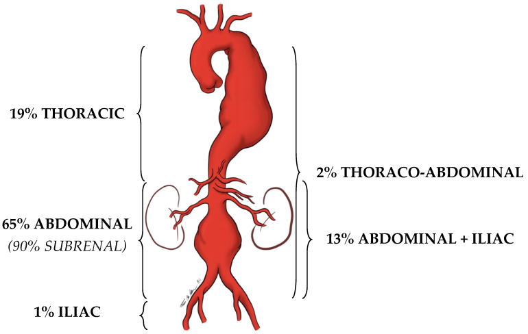
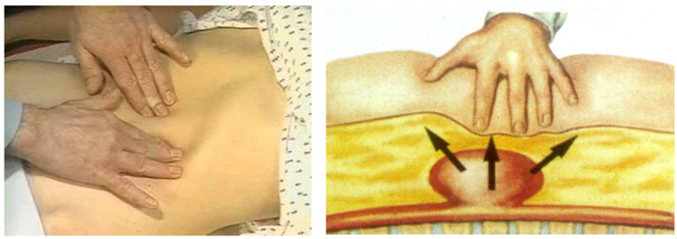
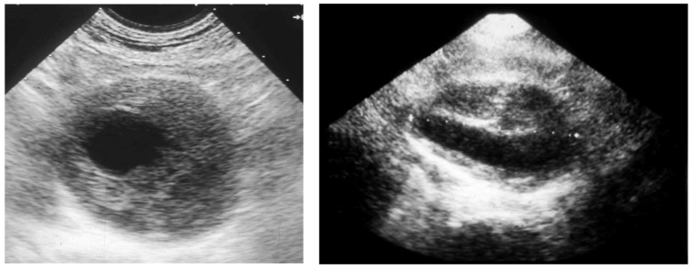
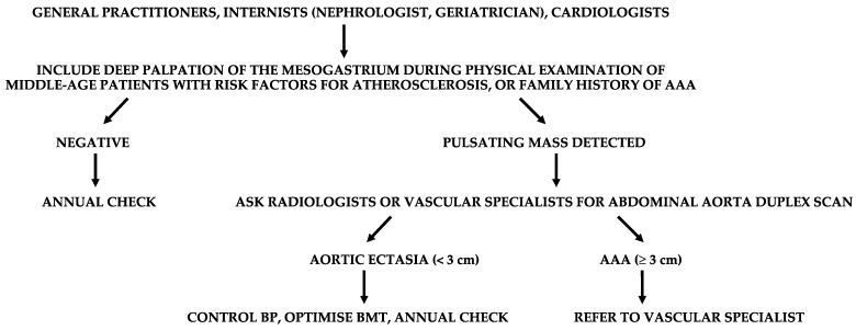
Middle-aged adults can start to be affected by some arterial diseases (ADs), such as abdominal aortic or popliteal artery aneurysms, lower extremity arterial disease, internal carotid, or renal artery or subclavian artery stenosis. These vasculopathies are often asymptomatic or paucisymptomatic before manifesting themselves with dramatic complications. Therefore, early detection of ADs is fundamental to reduce the risk of major adverse cardiovascular and limb events. Furthermore, ADs carry a high correlation with silent coronary artery disease (CAD). This study focuses on the most common ADs, in the attempt to summarize some key points which should selectively drive screening. Since the human and economic possibilities to instrumentally screen wide populations is not evident, deep knowledge of semeiotics and careful anamnesis must play a central role in our daily activity as physicians. The presence of some risk factors for atherosclerosis, or an already known history of CAD, can raise the clinical suspicion of ADs after a careful clinical history and a deep physical examination. The clinical suspicion must then be confirmed by a first-level ultrasound investigation and, if so, adequate treatments can be adopted to prevent dreadful complications.

Keywords: abdominal aortic aneurysm; carotid stenosis; complications; diagnosis; lower extremity arterial disease; popliteal artery aneurysm; renal artery stenosis; secondary prevention; subclavian artery stenosis.


Conflict of interest statement

The authors declare no conflict of interest.

Figures

Figure 1
Figure 1
Localization prevalence of aortic aneurysms.
Figure 2
Figure 2
Abdomen palpation in the search for an abdominal aortic aneurysm (AAA). An abnormal pulsating mass in the mesogastrium, expanding in all the directions, is strongly suspected to be an AAA.
Figure 3
Figure 3
Duplex scan of the abdominal aorta can easily confirm the clinical suspect of abdominal aortic aneurysm. Short axis (left) and long axis (right) view.
Figure 4
Figure 4
Suggested flow-chart for the detection of patients with abdominal aortic aneurysm (AAA). BP, blood pressure; BMT, best medical therapy.
Figure 5
Figure 5
Suggested flow-chart for the detection of patients with extracranial internal carotid stenosis (EICS). ATS, atherosclerosis; NASCET (North American Symptomatic Carotid Endarterectomy Trial criteria); BMT, best medical therapy.
Figure 6
Figure 6
Duplex scan (long axis view) of the extracranial carotid bifurcation, clearly showing an atherosclerotic plaque causing stenosis of the internal carotid artery.
Figure 7
Figure 7
Methods of measurement of severity of ICA stenosis, reproduced with permission from Bir and Kelley [43]. CCA, common carotid artery; ICA, internal carotid artery; ECA, external carotid artery; ECST, European Carotid Surgery Trial; NASCET, North American Symptomatic Carotid Endarterectomy Trial.
Figure 8
Figure 8
Arteriography showing obstruction of the left iliac axis, with an efficient collateral pathway revascularizing the common femoral artery. Depending on his/her age and lifestyle, this patient can eventually be asymptomatic or paucisymptomatic for intermittent claudication. An accurate physical examination (detecting the absence of the left femoral pulse), and the subsequent left ankle-brachial index (founded to be reduced) can easily allow for this patient to be classified as vasculopathic.
Figure 9
Figure 9
Suggested flow-chart for the detection of patients with lower extremity arterial disease (LEAD). IC, intermittent claudication; ATS, atherosclerosis; ABI, ankle-brachial index; BMT, best medical therapy.
Figure 10
Figure 10
On the left, the ankle-brachial index (ABI), an absolute number which inversely correlates with the severity of lower extremity arterial disease (from https://www.aemmedi.it/files/la_scuola_AMD/2014/diabete_e_arteriopatia/9a_SimoniIparte.pdf (accessed on 1 May 2023)). On the right, the ABI’s measurement technique (from Wikimedia: https://www.nhlbi.nih.gov/health/health-topics/topics/pad/diagnosis (accessed on 1 May 2023)).
Figure 11
Figure 11
Arteriography showing popliteal artery aneurysm. The physiologic flexion movement of the knee can dislocate part of the mural thrombus, which embolizes and occludes some tibial arteries, giving rise to clinical pictures ranging from an asymptomatic state to intermittent claudication, or chronic limb-threatening ischemia, or acute limb ischemia.
Figure 12
Figure 12
Suggested flow-chart for the detection of patients with popliteal artery aneurysm (PAA). ATS, atherosclerosis; AAA, abdominal aortic aneurysm; IC, intermittent claudication; ABI, ankle-brachial index; BP, blood pressure.
Figure 13
Figure 13
Suggested flow-chart for the detection of patients with renal artery stenosis (RAS). CRI, chronic renal insufficiency; BMT, best medical therapy.
Figure 14
Figure 14
Duplex scan of the renal arteries (long axis view). IVC, inferior vena cava.
Figure 15
Figure 15
Arteriography showing occlusion of the left prevertebral subclavian artery at its origin (on the left): the late angiogram (on the right) demonstrates the blood flow inversion in the left vertebral artery, revascularizing the left subclavian artery after the occlusion, that is subclavian steal syndrome.
Figure 16
Figure 16
Suggested flow-chart for the detection of patients with prevertebral subclavian artery stenosis (PSAS). BP, blood pressure. ATS, atherosclerosis. BMT, best medical therapy. CABG, coronary artery bypass graft.

References

    1. Johnston K.W., Rutherford R.B., Tilson M.D., Shah D.M., Hollier L., Stanley J.C. Suggested standards for reporting on arterial aneurysms. Subcommittee on Reporting Standards for Arterial Aneurysms, Ad Hoc Committee on Reporting Standards, Society for Vascular Surgery and North American Chapter, International Society for Cardiovascular Surgery. J. Vasc. Surg. 1991;13:452–458. doi: 10.1067/mva.1991.26737. - DOI - PubMed
    1. Chaikof E.L., Dalman R.L., Eskandari M.K., Jackson B.M., Lee W.A., Mansour M.A., Mastracci T.M., Mell M., Murad M.H., Nguyen L.L., et al. The Society for Vascular Surgery practice guidelines on the care of patients with an abdominal aortic aneurysm. J. Vasc. Surg. 2018;67:2–77. doi: 10.1016/j.jvs.2017.10.044. - DOI - PubMed
    1. Reimerink J.J., van der Laan M.J., Koelemay M.J., Balm R., Legemate D.A. Systematic review and meta-analysis of population-based mortality from ruptured abdominal aortic aneurysm. Br. J. Surg. 2013;100:1405–1413. doi: 10.1002/bjs.9235. - DOI - PubMed
    1. Kumar Y., Hooda K., Li S., Goyal P., Gupta N., Adeb M. Abdominal aortic aneurysm: Pictorial review of common appearances and complications. Ann. Transl. Med. 2017;5:256. doi: 10.21037/atm.2017.04.32. - DOI - PMC - PubMed
    1. Sakalihasan N., Michel J.B., Katsargyris A., Kuivaniemi H., Defraigne J.O., Nchimi A., Powell J.T., Yoshimura K., Hultgren R. Abdominal aortic aneurysms. Nat. Rev. Dis. Primers. 2018;4:34. doi: 10.1038/s41572-018-0030-7. - DOI - PubMed

LinkOut - more resources