Review
. 2023 Jul 14;13(14):2371.
doi: 10.3390/diagnostics13142371.

Surgical Anatomy of the Liver-Significance in Ovarian Cancer Surgery

Affiliations
Review

Surgical Anatomy of the Liver-Significance in Ovarian Cancer Surgery

Stoyan Kostov et al. Diagnostics (Basel). .

Abstract

Introduction: Ovarian cancer is the leading cause of death among all gynecological malignancies. Most patients present with an advanced stage of the disease. The routes of spread in ovarian cancer include peritoneal dissemination, direct invasion, and lymphatic or hematogenous spread, with peritoneal and lymphatic spread being the most common among them. The flow direction of the peritoneal fluid makes the right subphrenic space a target site for peritoneal metastases, and the most frequently affected anatomical area in advanced cases is the right upper quadrant. Complete cytoreduction with no macroscopically visible disease is the most important prognostic factor.

Methods: We reviewed published clinical anatomy reports associated with surgery of the liver in cases of advanced ovarian cancer.

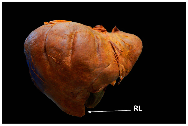
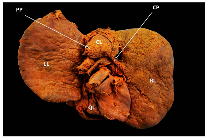
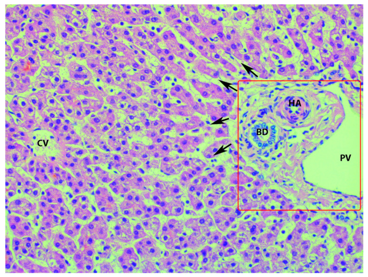
Results: The disease could disseminate anatomical areas, where complex surgery is required-Morrison's pouch, the liver surface, or porta hepatis. The aim of the present article is to emphasize and delineate the gross anatomy of the liver and its surgical application for oncogynecologists. Moreover, the association between the gross and microscopic anatomy of the liver is discussed. Additionally, the vascular supply and variations of the liver are clearly described.

Conclusions: Oncogynecologists performing liver mobilization, diaphragmatic stripping, and porta hepatis dissection must have a thorough knowledge of liver anatomy, including morphology, variations, functional status, potential diagnostic imaging mistakes, and anatomical limits of dissection.

Keywords: anatomical variations; hepatic veins; liver anatomy; liver ligaments; liver morphology; ovarian cancer surgery.


Conflict of interest statement

The authors declare no conflict of interest.

Figures

Figure 1
Figure 1
Different gross shapes of the liver—anterior surface of the liver (author’s own material): (A) globular shape; (B) conical shape of the right liver lobe and the small left lobe; (C) quadrilateral shape; (D) rectangle shape.
Figure 2
Figure 2
Morphological variations of the liver according to Netter’s classification (modified from references [18,19]). Type I—small left liver lobe and deep costal impression; Type II—left lobe atrophy; Type III—saddle-like liver with hypertrophied left lobe; Type IV—Riedel’s lobe; Type V—deep renal impression and corset construction; Type VI—diaphragmatic grooves.
Figure 3
Figure 3
Deep diaphragmatic liver grooves and liver fissures (author’s own material). (A)The arrows show two diaphragmatic grooves on the right liver lobe (anterior liver surface). (B) The arrows show three diaphragmatic grooves on the right liver lobe (anterior liver surface). (C) The arrow shows liver fissure on the right lobe (posterior liver surface). (D) The arrows show two liver fissures on the right lobe (posterior liver surface).
Figure 4
Figure 4
Riedel’s lobe of the liver—anterior liver surface (author’s own material). RL—Riedel’s lobe.
Figure 5
Figure 5
Riedel’s lobe on CT (author’s own material). RL—Riedel’s lobe.
Figure 6
Figure 6
Elongated caudate process and hypertrophied papillary process showed by arrows—posterior liver surface (own material). LL—left lobe; RL—right lobe; QL—quadrate lobe; CL— caudate lobe; CP—caudate process; PP—papillary process.
Figure 7
Figure 7
Unenhanced transverse abdominal CT liver level with isolated caudate lobe hypertrophy (own material). The arrow shows isolated caudate lobe hypertrophy.
Figure 8
Figure 8
Elongated papillary process (author’s own material). (A) Transverse abdominal CT at the level of the liver with elongated papillary process present. (B) Coronal abdominal CT where papillary liver process projection can be seen just above the portal vein. (C) CT Volume rendered image of the abdomen showing the papillary liver process in its typical location. Arrows show elongated papillary process.
Figure 9
Figure 9
Complete pons hepatis—type III (posterior liver surface). The fissure for ligamentum teres is absent (own material). LL—left liver lobe; RL—right liver lobe; FLV—fissure for ligamentum venosum; IVC—inferior vena cava; QL—quadrate lobe; CL—caudate lobe; GB—gall bladder; LTH—ligamentum teres hepatis; PH—pons hepatis.
Figure 10
Figure 10
Ligaments on the posterior (visceral) surface of the liver and porta hepatis (author’s own material). LL—left liver lobe; RL—right liver lobe; CL—caudate lobe; QL—quadrate lobe; GB—gall bladder; IVC—inferior vena cava; FLT—fissure for ligamentum teres; LT—ligamentum teres; LHA—left hepatic artery; RHA—right hepatic artery; PHA—proper hepatic artery; HPV—hepatic portal vein; CD—cystic duct; CHD—common hepatic duct; PH—porta hepatis.
Figure 11
Figure 11
Superior surface of the liver—part of the diaphragmatic surface (author’s own material). LL—left liver lobe; RL—right liver lobe; CL—caudate lobe; GB—gall bladder; AL—Arantius’ ligament; ML—Makuuchi ligament; ALVL—anterior layer of the coronary ligament on the right liver lobe; PLCL—posterior layer of the coronary ligament on the right liver lobe; TL—right triangular ligament; BA—bare area.
Figure 12
Figure 12
Liver ligament and their relationship with the lesser sac (omental bursa) (the author’s own material)—inferior and posterior surface of the liver. RL—right liver lobe; LL—left liver lobe; CL—caudal lobe; QL—quadrate lobe; GB—gall bladder; IFL—incised falciform ligament; FLT—fissure for ligamentum teres; LV—ligamentum venosum; LIPA—left inferior phrenic artery; LGA—left gastric artery; SA—splenic artery; CHA—common hepatic artery; SV—splenic vein; SMA—superior mesenteric artery; GDA—gastroduodenal artery; PV—portal vein; PHA—proper hepatic artery; LHA—left hepatic artery; CBD—common bile duct.
Figure 13
Figure 13
Attachments of coronary ligament and right triangular ligament (from the cadaveric dissection—archive of Dr. Ilker Selcuk). IVC—inferior vena cava.
Figure 14
Figure 14
Anatomy of the gastroduodenal ligament (from the cadaveric dissection—archive of Dr. Ilker Selcuk).
Figure 15
Figure 15
Segments of the liver according to Couinaud’s classification (own material): (A) ex vivo appearance; (B) in vivo appearance; (C) coronal volume-rendered abdominal CT image with frontal plane cut showing the annotated right hepatic lobe segment. Arabic and Latin numbers represents liver segment according to Couinaud’s classification. PV – portal vein; IVC – inferior vena cava.
Figure 16
Figure 16
Truncus celiacus and arterial supply of the liver (the author’s own material)—inferior and posterior surface of the liver. CL—caudate lobe; LL—left lobe; CTC—celiac trunk; SA—splenic artery; LGA—left gastric artery; LIPA—left inferior phrenic artery; RIPA—right inferior phrenic artery; CHA—common hepatic artery; GDA—gastroduodenal artery; HAP—proper hepatic artery; PV—portal vein; CBD—common bile duct.
Figure 17
Figure 17
Gross anatomy of the portal vein. The stomach and pancreas are retracted caudally to better visualize the tributaries of the portal vein (author’s own material). RL—right lobe of liver; ST—stomach; PC—pancreas; CHA—common hepatic artery; GDA—gastroduodenal artery; PV—portal vein; SMV—superior mesenteric vein; IMV—inferior mesenteric vein; SV—splenic vein.
Figure 18
Figure 18
Gross anatomy of hepatic veins—superior surface of the liver (author’s own material). LL—left liver lobe; RL—right liver lobe; IVC—inferior vena cava; LHV—left hepatic vein; MHV—middle hepatic vein; RHV—right hepatic vein.
Figure 19
Figure 19
Right and left hepatic vein (from the cadaveric dissection—archive of Dr. Ilker Selcuk).
Figure 20
Figure 20
Microanatomy of the liver lobule (author’s own material). PV—branch of portal vein; BD—branch of the bile duct; HA—branch of hepatic artery; CV—central vein; arrows—sinusoids; red square—the portal triad.
Figure 21
Figure 21
Sappey’s and Burrow’s veins ( adapted with permission from reference [63]). LTH—ligamentum teres hepatis; FL—falciform ligament; LPV—left portal vein; IEVs—inferior epigastric veins.
Figure 22
Figure 22
The white ellipse shows the narrow distance between the base of the ligamentum teres hepatis and the left branch of the portal vein (author’s own material). LTH—ligamentum teres hepatis; LL—left liver lobe; RL—right liver lobe; CL—caudate lobe; GB—gallbladder; PV—portal vein; RB—right branch of the portal vein; LB—left branch of the portal vein.
Figure 23
Figure 23
Base of the ligamentum teres hepatis (author’s own material—open surgery). (A) Peritoneal tumor dissemination of the base of the ligamentum teres hepatis in case of advanced ovarian cancer. (B) Removal of macroscopic peritoneal metastases at the base of the ligament. The round ligament of the liver was entirely removed. LTH—ligamentum teres hepatis, LL—left lobe of the liver; RL—right lobe of the liver; QL—quadrate lobe; LTH—ligamentum teres hepatis, GB—gall bladder.
Figure 24
Figure 24
Coronal post-contrast CT image of the abdomen, showing two accessory right inferior hepatic veins of the liver (author’s own material). RHV—right hepatic vein; AIRHV—accessory inferior right hepatic vein.
Figure 25
Figure 25
Limits of the bare area of the liver on the diaphragmatic surface (own material). (The liver was removed for better identification.) AN—area nuda; RAg—right adrenal gland; RKi—right kidney.
Figure 26
Figure 26
Small retrohepatic additional right liver lobe vein, which drains directly into the IVC—visceral surface of the liver (author’s own material). IVC—inferior vena cava; AV— additional vein; LL—left liver lobe; RL—right liver lobe.
Figure 27
Figure 27
Some steps of liver mobilization followed by diaphragmatic stripping (author’s own material). (A) Step 1—dissection of falciform ligament (embalmed cadaver). (B,C) Steps 6,7, 8—dissection of posterior layer of the coronary ligament on the right liver lobe and dissection of the right triangular ligament. (C) Final view after diaphragmatic stripping in case of massive metastases on the right diaphragmatic peritoneum ((BD) open surgery). ACL—anterior layer of the coronary ligament; RL—right lobe of the liver; LL—left lobe of the liver; FL—falciform ligament; PCL—posterior layer of the coronary ligament on the right liver lobe; RTL—right triangular ligament.
Figure 28
Figure 28
Kocher (AC) and Pringle (D) maneuvers (embalmed cadavers—author’s own material). (A) Incision of the lateral peritoneum, which started between the lateral aspect of the epiploic foramen and the inferior duodenal flexure. (B) Dissection of fascia of Treitz, which is located below the head of the pancreas. (C) Exposure of the inferior vena cava until the medial limit of dissection—the left renal vein. (D) Vessel loop, which encircles the hepatoduodenal ligament. HP—head of the pancreas, D—duodenum, GB—gall bladder; K— kidney; PV—portal vein, LRV—left renal vein; prop IVC—inferior vena cava; PHA—proper hepatic artery; CBD—common bile duct; LL—left lobe of the liver.

References

    1. Taskiran C., Vatansever D., Giray B., Eraslan A., Tanju S., Arvas M., Balik E. Upper abdominal debulking surgery for ovarian cancer total colectomy, total peritonectomy, and extended upper abdominal debulking surgery. Int. J. Gynecol. Cancer. 2020;30:1648–1649. doi: 10.1136/ijgc-2020-001531. - DOI - PubMed
    1. Aletti G.D., Dowdy S.C., Gostout B.S., Jones M.B., Stanhope C.R., Wilson T.O., Podratz K.C., Cliby W.A. Aggressive surgical effort and improved survival in advanced-stage ovarian cancer. Obs. Gynecol. 2006;107:77–85. doi: 10.1097/01.AOG.0000192407.04428.bb. - DOI - PubMed
    1. Bristow R.E., Karlan B.Y., Chi D.S. Surgery for Ovarian Cancer. 3rd ed. Taylor & Francis Group, LLC.; Abingdon, UK: 2016.
    1. Pannu H.K., Oliphant M. The subperitoneal space and peritoneal cavity: Basic concepts. Abdom. Imaging. 2015;40:2710–2722. doi: 10.1007/s00261-015-0429-5. - DOI - PMC - PubMed
    1. Stepanyan A., Malakyan Z., Alaverdyan A., Davtyan H., Hovhannisyan T. Right upper quadrant peritonectomy. Answering frequently asked questions. Int. J. Gynecol. Cancer. 2021;31:1305–1306. doi: 10.1136/ijgc-2021-002695. - DOI - PubMed

LinkOut - more resources