. 2023 Jul 20;13(14):2426.
doi: 10.3390/diagnostics13142426.

Machine-Learning-Based Diagnostics of Cardiac Sarcoidosis Using Multi-Chamber Wall Motion Analyses

Affiliations

Machine-Learning-Based Diagnostics of Cardiac Sarcoidosis Using Multi-Chamber Wall Motion Analyses

Jan Eckstein et al. Diagnostics (Basel). .

Abstract

Background: Hindered by its unspecific clinical and phenotypical presentation, cardiac sarcoidosis (CS) remains a challenging diagnosis.

Objective: Utilizing cardiac magnetic resonance imaging (CMR), we acquired multi-chamber volumetrics and strain feature tracking for a support vector machine learning (SVM)-based diagnostic approach to CS.

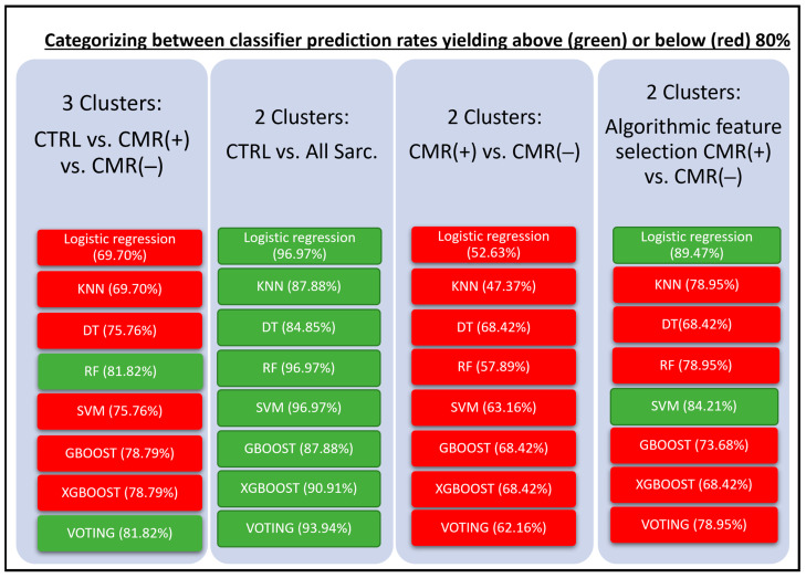
Method: Forty-five CMR-negative (CMR(-), 56.5(53.0;63.0)years), eighteen CMR-positive (CMR(+), 64.0(57.8;67.0)years) sarcoidosis patients and forty-four controls (CTRL, 56.5(53.0;63.0)years)) underwent CMR examination. Cardiac parameters were processed using the classifiers of logistic regression, KNN(K-nearest-neighbor), DT (decision tree), RF (random forest), SVM, GBoost, XGBoost, Voting and feature selection.

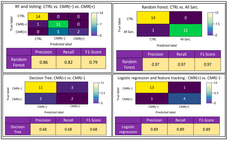
Results: In a three-cluster analysis of CTRL versus vs. CMR(+) vs. CMR(-), RF and Voting classifier yielded the highest prediction rates (81.82%). The two-cluster analysis of CTRL vs. all sarcoidosis (All Sarc.) yielded high prediction rates with the classifiers logistic regression, RF and SVM (96.97%), and low prediction rates for the analysis of CMR(+) vs. CMR(-), which were augmented using feature selection with logistic regression (89.47%).

Conclusion: Multi-chamber cardiac function and strain-based supervised machine learning provides a non-contrast approach to accurately differentiate between healthy individuals and sarcoidosis patients. Feature selection overcomes the algorithmically challenging discrimination between CMR(+) and CMR(-) patients, yielding high accuracy predictions. The study findings imply higher prevalence of cardiac involvement than previously anticipated, which may impact clinical disease management.

Keywords: cardiac magnetic resonance; cardiac sarcoidosis 3; cardiac strain; machine learning 2; multi-chamber analyses.


Conflict of interest statement

The authors declare no conflict of interest.

Figures

Figure 1
Figure 1
Flow chart illustrating the processing steps of machine learning along with the applied classifiers. CTRL—control subjects, CMR(+)—positive cardiac magnetic resonance patients, CMR(−)—negative cardiac magnetic resonance patients, KNN—K-nearest-neighbor, DT—deep trees, RF—random forest, SVM—support vector machine, GBOOST—gradient boosting, XGBOOST—extreme gradient boosting.
Figure 2
Figure 2
Graphic discrimination between classifier performance of the different analyses, yielding below (red) and above (green) 80% prediction rates with corresponding outcome statistics. CTRL—control subjects, CMR(+)—positive cardiac magnetic resonance patients, CMR(−)—negative cardiac magnetic resonance patients, All Sarc.—all sarcoidosis patients, KNN—K-nearest-neighbor, DT—deep trees, RF—random forest, SVM—support vector machine, GBOOST—gradient boosting, XGBOOST—extreme gradient boosting.
Figure 3
Figure 3
Selected confusion matrices for 36-feature analyses of the various cluster analyses, depicting the highest yielding classifier(s) per cluster with corresponding performance statistics. Precision—positive predictive value, recall score—sensitivity, F1-score—test accuracy, CTRL—control subjects, CMR(+)—positive cardiac magnetic resonance patients, CMR(−)—negative cardiac magnetic resonance patients, All Sarc.—all sarcoidosis patients, DT—deep trees, RF—random forest.
Figure 4
Figure 4
ROC curves for (A) CMR(+) vs. CMR(–) with no feature selection and (B) (+) v. CMR(–) after selecting features.

References

    1. Soto-Gomez N., Peters J.I., Nambiar A.M. Diagnosis and Management of Sarcoidosis. Am. Fam. Physician. 2016;93:840–848. - PubMed
    1. Hamzeh N., Steckman D.A., Sauer W.H., Judson M.A. Pathophysiology and clinical management of cardiac sarcoidosis. Nat. Rev. Cardiol. 2015;12:278–288. doi: 10.1038/nrcardio.2015.22. - DOI - PubMed
    1. Chow K.L., O’Donnell J.L., Crozier I. Prevalence, incidence and survival outcomes of cardiac sarcoidosis in the South Island, New Zealand. Int. J. Cardiol. 2022;357:128–133. doi: 10.1016/j.ijcard.2022.04.004. - DOI - PubMed
    1. Swigris J.J., Olson A.L., Huie T.J., Fernandez-Perez E.R., Solomon J., Sprunger D., Brown K.K. Sarcoidosis-related mortality in the United States from 1988 to 2007. Am. J. Respir. Crit. Care Med. 2011;183:1524–1530. doi: 10.1164/rccm.201010-1679OC. - DOI - PMC - PubMed
    1. Silverman K.J., Hutchins G.M., Bulkley B.H. Cardiac sarcoid: A clinicopathologic study of 84 unselected patients with systemic sarcoidosis. Circulation. 1978;58:1204–1211. doi: 10.1161/01.CIR.58.6.1204. - DOI - PubMed

LinkOut - more resources