Review
. 2023 Jul 21;13(14):2440.
doi: 10.3390/diagnostics13142440.

Thoracic Diseases: Technique and Applications of Dual-Energy CT

Affiliations
Review

Thoracic Diseases: Technique and Applications of Dual-Energy CT

Armando Perrella et al. Diagnostics (Basel). .

Abstract

Dual-energy computed tomography (DECT) is one of the most promising technological innovations made in the field of imaging in recent years. Thanks to its ability to provide quantitative and reproducible data, and to improve radiologists' confidence, especially in the less experienced, its applications are increasing in number and variety. In thoracic diseases, DECT is able to provide well-known benefits, although many recent articles have sought to investigate new perspectives. This narrative review aims to provide the reader with an overview of the applications and advantages of DECT in thoracic diseases, focusing on the most recent innovations. The research process was conducted on the databases of Pubmed and Cochrane. The article is organized according to the anatomical district: the review will focus on pleural, lung parenchymal, breast, mediastinal, lymph nodes, vascular and skeletal applications of DECT. In conclusion, considering the new potential applications and the evidence reported in the latest papers, DECT is progressively entering the daily practice of radiologists, and by reading this simple narrative review, every radiologist will know the state of the art of DECT in thoracic diseases.

Keywords: ILD; acute aortic syndromes; breast cancer; dual-energy CT; esophageal cancer; lung cancer; lymph nodes; pleural carcinomatosis; pulmonary embolism; thymoma.


Conflict of interest statement

The authors declare no conflict of interest.

Figures

Figure 1
Figure 1
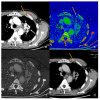
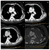
Qualitative evaluation of pleural carcinosis using an arterial phase with a conventional CT scan (a) and DECT scan with virtual monoenergetic reconstruction at 80 keV (b) and 40 keV (c). Low-energy monoenergetic reconstructions provide better conspicuity of the pleural lesion. The white arrow indicates the pleural metastasis.
Figure 2
Figure 2
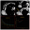
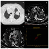
Evaluation with 40 keV monoenergetic reconstruction (a) and iodine map (b) of a pleural lesion (yellow arrow) found in the CT scan of a patient with hilar lung neoplasia (a white arrow). The pleural lesion has an iodine concentration of 1.2 mg/mL (c), which is therefore compatible with a benign lesion [46]. The benignity of the lesion is demonstrated by the spontaneous regression of the lesion after 2 months (d).
Figure 3
Figure 3
Evaluation of pleural carcinomatosis (white arrow) secondary to clear cell renal cancer with monoenergetic reconstruction at 40 keV (a) and iodine map (b). The iodine concentration is 5.2 mg/mL (c), thus indicative of the malignancy of the finding [46].
Figure 4
Figure 4
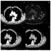
Assessment of breast neoplasia (white arrows) with monoenergetic reconstructions at 80 keV (a,d), 40 keV (b,e), and iodine map (c,f). Low monoenergetic reconstructions and iodine maps provide the highest conspicuity.
Figure 5
Figure 5
In a case of breast cancer (a), DECT analysis allows us to distinguish between a normal lymph node ((b), white arrow) and a pathological lymph node ((c), yellow arrow). In fact, the slope HU values (d) of a pathological lymph node (red ROI) and breast cancer (blue ROI) were similar if compared to normal lymph node (yellow ROI). In iodine concentration evaluation with scatterplot (e), there is a complete separation between breast cancer (blue ROI) and pathological lymph node (red ROI) compared to normal lymph node (yellow ROI).
Figure 6
Figure 6
In a case of breast cancer (a), DECT analysis allows for distinguishing between a normal mediastinal lymph node ((a), green arrow and green ROI) and metastatic axillary and internal mammary lymph nodes ((b), yellow arrow and yellow ROI). Pathological lymph nodes present a slope of spectral HU curve (c) perfectly matching that of breast cancer ((a), white arrow, blue ROI).
Figure 7
Figure 7
A case of spindle cell thymoma, type A, stage IIB according to Masaoka ((a), white arrow) with an iodine concentration of 1.22 mg/dL (b), and a density at 60 keV of 73.12 HU (c) and at 70 keV of 60.97 HU (d).
Figure 8
Figure 8
A case of thymic basaloid carcinoma ((a), white arrow). The iodine concentration is 3.4 mg/mL (b), the density at 60 keV is 139.47 HU (c), and at 70 keV is 100.31 HU (d).
Figure 9
Figure 9
DECT provides the characterization of a thymic cyst ((a) white arrow). The density at 60 keV is 25.93 HU (b), while that at 70 keV is 18.60 HU (c), and the iodine concentration is 0.85 mg/dL (d). All values are below the cut-offs (according to Zhou et al.) to distinguish it from hypovascular solid tissue [97].
Figure 10
Figure 10
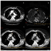
A case of esophageal adenocarcinoma with extension beyond the gastro-esophageal junction, evaluated with conventional CT in the arterial phase (a) and DECT in the delayed phase with iodine map (b) and monoenergetic reconstructions at 60 keV (c) and 40 keV (d).
Figure 11
Figure 11
Case of aortic intramural hematoma (a), in which true non-contrast (b) and virtual non-contrast (c) both demonstrate efficacy in detecting intrinsic hyperintensity of the hematoma.
Figure 12
Figure 12
DECT allows increased confidence in detecting the presence of pulmonary embolism (yellow arrows) thanks to the increased contrast resolution provided by low-energy VMI (a), but it also provides the opportunity to assess the parenchymal distribution of iodine by identifying the corresponding lung perfusion defects on the iodine map ((b) white arrows).
Figure 13
Figure 13
In this patient with lung cancer (a), neoplastic thrombosis is documented in the right atrium. The diagnosis is confirmed by the evidence of iodine concentration in both the primary neoplasm (b) and the thrombotic formation (c). The analysis is performed by scatterplot (d) yellow ROI neoplasm and red ROI neoplastic thrombus present a similar pathologic iodine concentration).
Figure 14
Figure 14
Spectral curve analysis allows a distinction to be made between osteosclerotic metastases ((a,b) pink, blue and light blue ROIs) and porotic vertebral fracture ((a,b) red and yellow ROIs). The spectral curve (c) demonstrates a perfect separation of the attenuation profiles between the two findings.
Figure 15
Figure 15
The iodine map (b) increases the visibility of a metastasis of the left clavicle compared to conventional CT scan with polychromatic beam (a).

References

    1. Johnson T.R.C., Krauss B., Sedlmair M., Grasruck M., Bruder H., Morhard D., Fink C., Weckbach S., Lenhard M., Schmidt B., et al. Material differentiation by dual energy CT: Initial experience. Eur. Radiol. 2007;17:1510–1517. doi: 10.1007/s00330-006-0517-6. - DOI - PubMed
    1. Agostini A., Borgheresi A., Mari A., Floridi C., Bruno F., Carotti M., Schicchi N., Barile A., Maggi S., Giovagnoni A. Dual-energy CT: Theoretical principles and clinical applications. Radiol. Med. 2019;124:1281–1295. doi: 10.1007/s11547-019-01107-8. - DOI - PubMed
    1. Carotti M., Salaffi F., Beci G., Giovagnoni A. The application of dual-energy computed tomography in the diagnosis of musculoskeletal disorders: A review of current concepts and applications. Radiol. Med. 2019;124:1175–1183. doi: 10.1007/s11547-019-01015-x. - DOI - PubMed
    1. Biondi M., Vanzi E., De Otto G., Buonamici F.B., Belmonte G., Mazzoni L., Guasti A., Carbone S.F., Mazzei M.A., La Penna A., et al. Water/cortical bone decomposition: A new approach in dual energy CT imaging for bone marrow oedema detection. A feasibility study. Phys. Medica. 2016;32:1712–1716. doi: 10.1016/j.ejmp.2016.08.004. - DOI - PubMed
    1. Foley W.D., Shuman W.P., Siegel M.J. White Paper of the Society of Computed Body Tomography and Magnetic Resonance on Dual-Energy CT, Part 2: Radiation Dose and Iodine Sensitivity. J. Comput. Assist Tomogr. 2016;40:846–850. doi: 10.1097/RCT.0000000000000539. - DOI - PubMed

LinkOut - more resources