

Skin Bank Establishment in Treatment of Severe Burn Injuries: Overview and Experience with Skin Allografts at the Vienna Burn Center
- PMID: 37510832
- PMCID: PMC10381394
- DOI: 10.3390/jcm12144717
Skin Bank Establishment in Treatment of Severe Burn Injuries: Overview and Experience with Skin Allografts at the Vienna Burn Center
Abstract
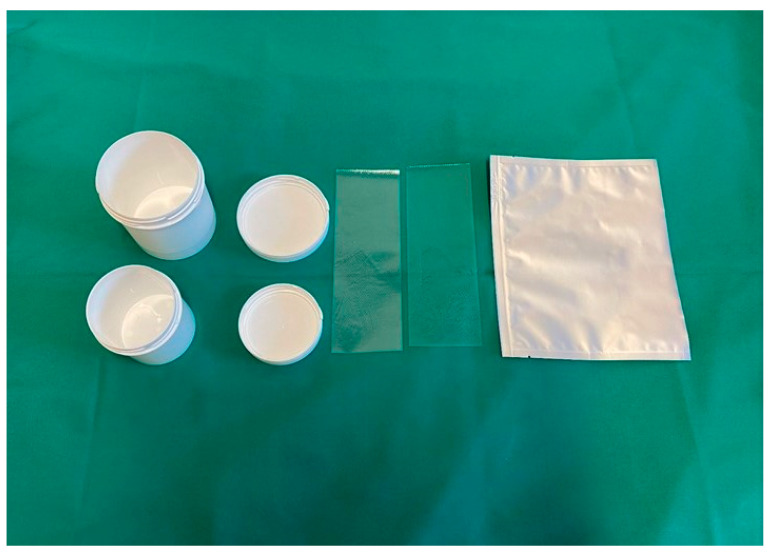
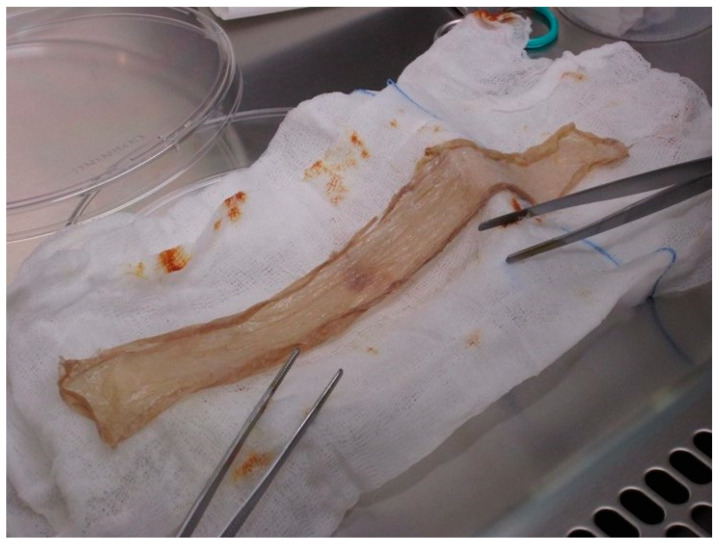
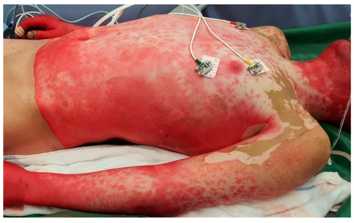
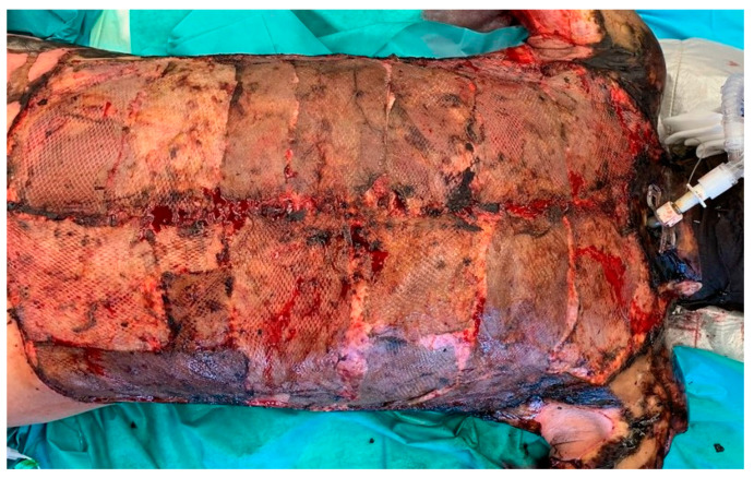
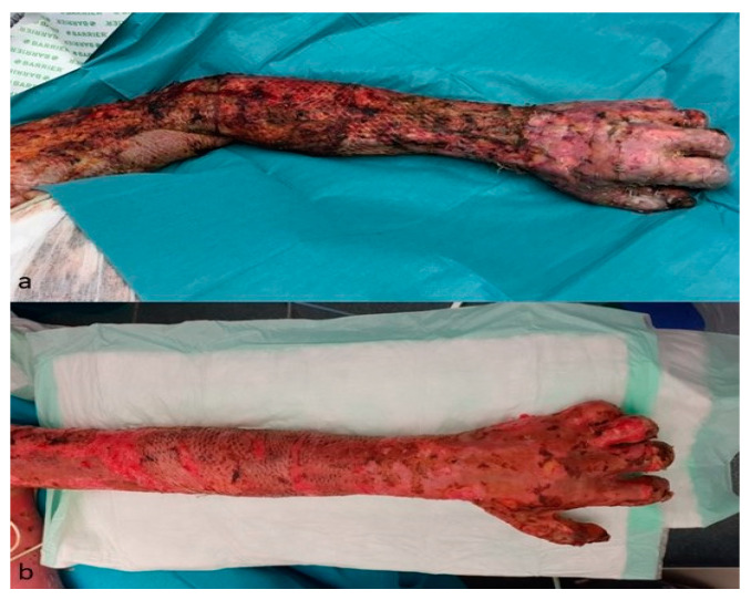
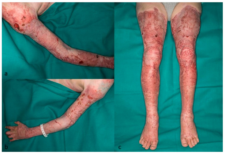
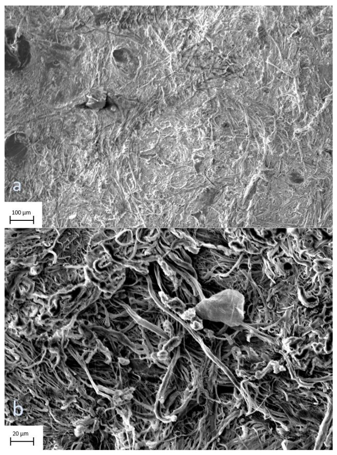
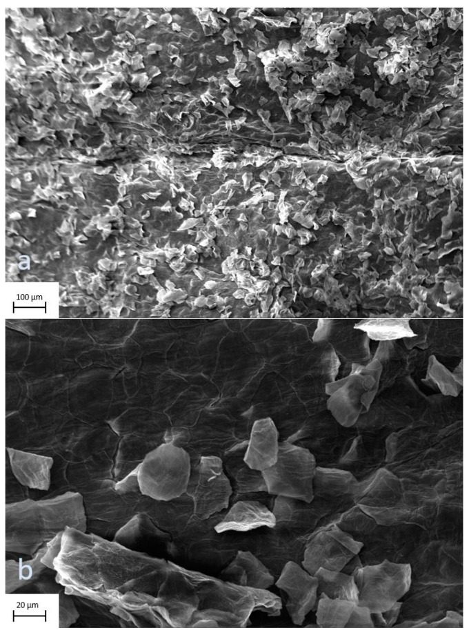
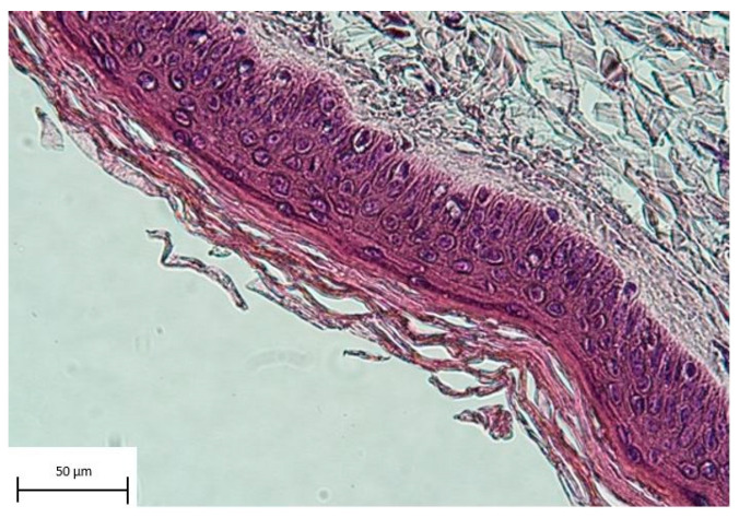
Depending on their extent, burn injuries require different treatment strategies. In cases of severe large-area trauma, the availability of vital skin for autografting is limited. Donor skin allografts are a well-established but rarely standardized option for temporary wound coverage. Ten patients were eligible for inclusion in this retrospective study. Overall, 202 donor skin grafts obtained from the in-house skin bank were applied in the Department of Plastic and Reconstructive and Aesthetic Surgery, Medical University of Vienna. Between 2017 and 2022, we analysed the results in patient treatment, the selection of skin donors, tissue procurement, tissue processing and storage of allografts, as well as the condition and morphology of the allografts before application. The average Abbreviated Burn Severity Index (ABSI) was 8.5 (range, 5-12), and the mean affected total body surface area (TBSA) was 46.1% (range, 20-80%). In total, allograft application was performed 14 times. In two cases, a total of eight allografts were removed due to local infection, accounting for 3.96% of skin grafts. Six patients survived the acute phase of treatment. Scanning electron microscope images and histology showed no signs of scaffold decomposition and intact tissue layers of the allografts. The skin banking program and the application of skin allografts at the Vienna Burn Center can be considered successful. In severe burn injuries, skin allografts provide time by serving as sufficient wound coverage after early necrosectomy. Having an in-house skin banking program at a dedicated burn centre is particularly advantageous since issues of availability and distribution can be minimized. Skin allografts provide a reliable treatment option in patients with extensive burn injuries.
Keywords: burn wound; skin allograft; skin bank; skin regeneration; skin substitute.
Conflict of interest statement
The authors declare no conflict of interest.
Figures
Similar articles
-
26 Years of Skin Banking in Finland.Eur Burn J. 2024 Dec 9;5(4):429-437. doi: 10.3390/ebj5040038. Eur Burn J. 2024. PMID: 39727914 Free PMC article.
-
A Study of Cadaveric Skin Graft Harvest and Usage: An Observational Prospective Pilot Study.Cureus. 2024 Sep 22;16(9):e69932. doi: 10.7759/cureus.69932. eCollection 2024 Sep. Cureus. 2024. PMID: 39439632 Free PMC article.
-
Experience and outcomes of micrografting for major paediatric burns.Burns. 2017 Aug;43(5):1103-1110. doi: 10.1016/j.burns.2017.02.008. Epub 2017 Mar 18. Burns. 2017. PMID: 28318749
-
Glycerolised Skin Allografts for Extensive Burns in Low- and Middle-income Countries.J West Afr Coll Surg. 2021 Jul-Sep;11(3):35-41. doi: 10.4103/jwas.jwas_55_21. Epub 2022 Jul 22. J West Afr Coll Surg. 2021. PMID: 36132972 Free PMC article. Review.
-
Clinical Applications of Allograft Skin in Burn Care.Ann Plast Surg. 2020 Mar;84(3S Suppl 2):S158-S160. doi: 10.1097/SAP.0000000000002282. Ann Plast Surg. 2020. PMID: 32028339 Review.
Cited by
-
Synergistic Treatment of Infected Burn Wound Utilizing Maggot Debridement and Acellular Fish Skin Grafting-A Case Report.J Burn Care Res. 2024 Sep 6;45(5):1336-1340. doi: 10.1093/jbcr/irae128. J Burn Care Res. 2024. PMID: 38953562 Free PMC article.
-
Double Capsule Phenomenon After Breast Implant Exchange: Safety and Technical Considerations.Aesthetic Plast Surg. 2025 Feb;49(3):1017-1018. doi: 10.1007/s00266-023-03808-y. Epub 2023 Dec 30. Aesthetic Plast Surg. 2025. PMID: 38165404 No abstract available.
-
26 Years of Skin Banking in Finland.Eur Burn J. 2024 Dec 9;5(4):429-437. doi: 10.3390/ebj5040038. Eur Burn J. 2024. PMID: 39727914 Free PMC article.
-
Current State and Challenges of Tissue and Organ Cryopreservation in Biobanking.Int J Mol Sci. 2024 Oct 16;25(20):11124. doi: 10.3390/ijms252011124. Int J Mol Sci. 2024. PMID: 39456905 Free PMC article. Review.
-
An Overview of Recent Developments in the Management of Burn Injuries.Int J Mol Sci. 2023 Nov 15;24(22):16357. doi: 10.3390/ijms242216357. Int J Mol Sci. 2023. PMID: 38003548 Free PMC article. Review.
References
-
- Tam J., Wang Y., Farinelli W.A., Jimenez-Lozano J., Franco W., Sakamoto F.H., Cheung E.J., Purschke M., Doukas A.G., Anderson R.R. Fractional Skin Harvesting: Autologous Skin Grafting without Donor-site Morbidity. Plast. Reconstr. Surg. Glob. Open. 2013;1:e47. doi: 10.1097/GOX.0b013e3182a85a36. - DOI - PMC - PubMed
LinkOut - more resources
Full Text Sources
