Review
. 2023 Jul 24;12(14):4866.
doi: 10.3390/jcm12144866.

The Role of Multimodality Imaging in Pediatric Cardiomyopathies

Affiliations
Review

The Role of Multimodality Imaging in Pediatric Cardiomyopathies

Sara Moscatelli et al. J Clin Med. .

Abstract

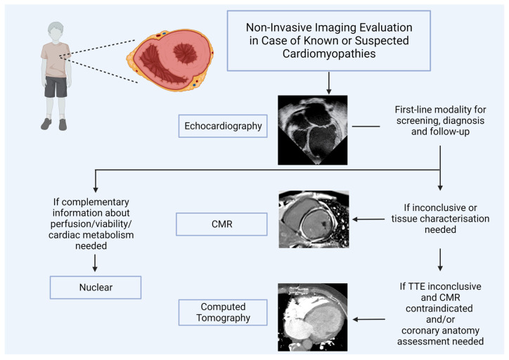
Cardiomyopathies are a heterogeneous group of myocardial diseases representing the first cause of heart transplantation in children. Diagnosing and classifying the different phenotypes can be challenging, particularly in this age group, where cardiomyopathies are often overlooked until the onset of severe symptoms. Cardiovascular imaging is crucial in the diagnostic pathway, from screening to classification and follow-up assessment. Several imaging modalities have been proven to be helpful in this field, with echocardiography undoubtedly representing the first imaging approach due to its low cost, lack of radiation, and wide availability. However, particularly in this clinical context, echocardiography may not be able to differentiate from cardiomyopathies with similar phenotypes and is often complemented with cardiovascular magnetic resonance. The latter allows a radiation-free differentiation between different phenotypes with unique myocardial tissue characterization, thus identifying the presence and extent of myocardial fibrosis. Nuclear imaging and computed tomography have a complementary role, although they are less used in daily clinical practice due to the concern related to the use of radiation in pediatric patients. However, these modalities may have some advantages in evaluating children with cardiomyopathies. This paper aims to review the strengths and limitations of each imaging modality in evaluating pediatric patients with suspected or known cardiomyopathies.

Keywords: cardiovascular multimodality imaging; pediatric cardiology; pediatric cardiomyopathy.


Conflict of interest statement

The authors declare no conflict of interest.

Figures

Figure 1
Figure 1
(a) Parasternal short-axis view in a 14-year-old female with ventricular tachycardia during exercise, demonstrating right ventricular (RV) enlargement with suspected ACM. (b) TDI of the RV free-wall showing diminished tricuspid annular TD velocities of the same patient. (c) Parasternal long-axis view in a 13-year-old male with SIV hypertrophy in HCM. (d) Parasternal short-axis view showing hypertrophy of mid-ventricular IVS. (e) Apical-4-chamber view showing dilated LV and LA in suspected DCM. (f) Parasternal short-axis view demonstrates LVEDD at the papillary muscles level. The images (g,h) show two different cases of LVNC.
Figure 2
Figure 2
Cardiovascular Magnetic Resonance Imaging in three patients with different cardiomyopathy phenotypes. Panel (A): early hypertrophic cardiomyopathy (HCM). There is concentric left ventricular hypertrophy (left), accompanied by prolonged T1 mapping values (center) and no late gadolinium enhancement. The white asterisk (left) indicates the hypertrophic septum. Panel (B): dilated cardiomyopathy (DCM) in a patient with Duchenne muscular dystrophy. There is moderate chamber enlargement and LV systolic dysfunction (LVEF 37%) (left). T1 mapping values are globally elevated (center), and there is mainly epicardial late enhancement of the basal to mid lateral wall (right).
Figure 3
Figure 3
Cardiovascular computed tomography imaging presenting three patients with different cardiomyopathy. Panel (A): Hypertrophic Cardiomyopathy (HCM). The white asterisk indicates the hypertrophic septum. Panel (B): Arrhythmogenic cardiomyopathy right. The black arrows indicate the fibrofatty deposition of the right ventricle in the dual-energy CCT. Panel (C): Left ventricular non-compaction. The black asterisks indicate the prominent trabeculae of the left ventricle. Right ventricle (RV); left ventricle (LV); right atrium (RA); left atrium (LA).
Figure 4
Figure 4
Myocardial perfusion imaging with Single Photon Emission Computed Tomography (SPECT) in a pediatric patient: both stress and rest images (axial images in first and second lines, vertical long axis images in third and fourth lines, horizontal long axis images in fifth and sixth lines) show left ventricular posterior wall and septal hypertrophy [98].
Figure 5
Figure 5
Cardiovascular imaging algorithm in the pediatric cardiomyopathy field.

References

    1. Lee T.M., Hsu D.T., Kantor P., Towbin J.A., Ware S.M., Colan S.D., Chung W.K., Jefferies J.L., Rossano J.W., Castleberry C.D., et al. Pediatric Cardiomyopathies. Circ. Res. 2017;121:855–873. doi: 10.1161/CIRCRESAHA.116.309386. - DOI - PMC - PubMed
    1. Lipshultz S.E., Sleeper L.A., Towbin J.A., Lowe A.M., Orav E.J., Cox G.F., Lurie P.R., McCoy K.L., McDonald M.A., Messere J.E., et al. The Incidence of Pediatric Cardiomyopathy in Two Regions of the United States. N. Engl. J. Med. 2003;348:1647–1655. doi: 10.1056/NEJMoa021715. - DOI - PubMed
    1. Webber S.A. New-Onset Heart Failure in Children in the Absence of Structural Congenital Heart Disease. Circulation. 2008;117:11–12. doi: 10.1161/CIRCULATIONAHA.107.747469. - DOI - PubMed
    1. Nugent A.W., Daubeney P.E., Chondros P., Carlin J.B., Cheung M., Wilkinson L.C., Davis A.M., Kahler S.G., Chow C., Wilkinson J.L., et al. The Epidemiology of Childhood Cardiomyopathy in Australia. N. Engl. J. Med. 2003;348:1639–1646. doi: 10.1056/NEJMoa021737. - DOI - PubMed
    1. Lipshultz S.E., Law Y.M., Asante-Korang A., Austin E.D., Dipchand A.I., Everitt M.D., Hsu D.T., Lin K.Y., Price J.F., Wilkinson J.D., et al. Cardiomyopathy in Children: Classification and Diagnosis: A Scientific Statement from the American Heart Association. Circulation. 2019;140:e9–e68. doi: 10.1161/CIR.0000000000000682. - DOI - PubMed

LinkOut - more resources