. 2023 Jul 8;24(14):11252.
doi: 10.3390/ijms241411252.

A Novel Rat Model of ADHD-like Hyperactivity/Impulsivity after Delayed Reward Has Selective Loss of Dopaminergic Neurons in the Right Ventral Tegmental Area

Affiliations

A Novel Rat Model of ADHD-like Hyperactivity/Impulsivity after Delayed Reward Has Selective Loss of Dopaminergic Neurons in the Right Ventral Tegmental Area

Sarah E Kohe et al. Int J Mol Sci. .

Abstract

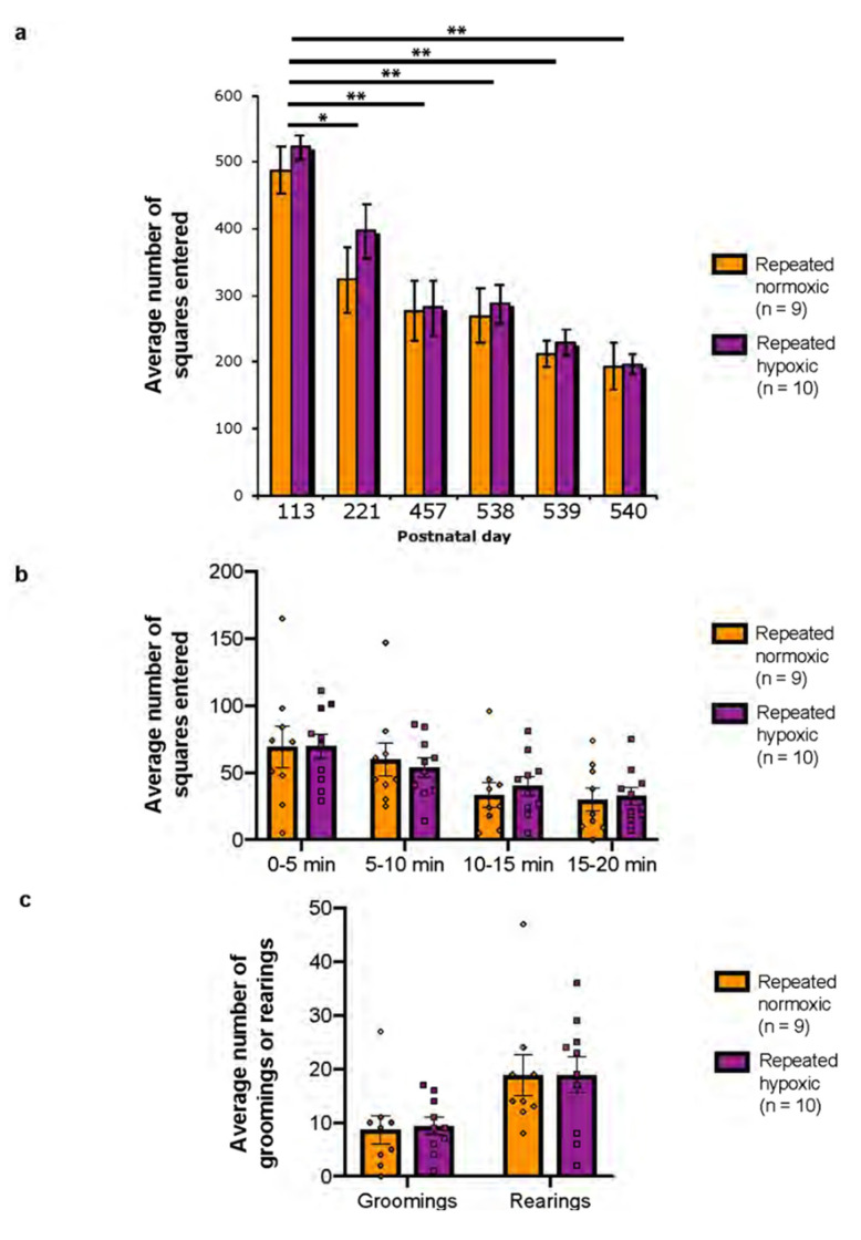
In attention deficit hyperactivity disorder (ADHD), hyperactivity and impulsivity occur in response to delayed reward. Herein we report a novel animal model in which male Sprague-Dawley rats exposed to repeated hypoxic brain injury during the equivalent of extreme prematurity were ADHD-like hyperactive/impulsive in response to delayed reward and attentive at 3 months of age. Thus, a unique animal model of one of the presentations/subtypes of ADHD was discovered. An additional finding is that the repeated hypoxia rats were not hyperactive in the widely used open field test, which is not ADHD specific. Hence, it is recommended that ADHD-like hyperactivity and ADHD-like impulsivity, specifically in response to delayed reward, be a primary component in the design of future experiments that characterize potential animal models of ADHD, replacing open field testing of hyperactivity. Unknown is whether death and/or activity of midbrain dopaminergic neurons contributed to the ADHD-like hyperactivity/impulsivity detected after delayed reward. Hence, we stereologically measured the absolute number of dopaminergic neurons in four midbrain subregions and the average somal/nuclear volume of those neurons. Repeated hypoxia rats had a significant specific loss of dopaminergic neurons in the right ventral tegmental area (VTA) at 2 weeks of age and 18 months of age, providing new evidence of a site of pathology. No dopaminergic neuronal loss occurred in three other midbrain regions. Fewer VTA dopaminergic neurons correlated with increased ADHD-like hyperactivity and impulsivity. Novel early intervention therapies to rescue VTA dopaminergic neurons and potentially prevent ADHD-like hyperactivity/impulsivity can now be investigated.

Keywords: ADHD hyperactive/impulsive presentation; fixed-interval extinction test; midbrain dopaminergic neurons; open field test; stereology; ventral tegmental area.


Conflict of interest statement

The authors declare no conflict of interest.

Figures

Figure 1
Figure 1
Timelines for the experiments. (a) Experimental timeline for the long-term cohort of repeated normoxia and repeated hypoxia rats (Cohort 1). This cohort was used for open field testing at 4, 7.5, 15, and 18 months of age, fixed interval–extinction (FI-EXT) testing from 16 months of age, and histochemical and immunohistochemical investigation of midbrain neurons. * Data from testing on the open field are shown in this paper. ** Hyperactivity and inattention data were published in Oorschot et al. [20]. Impulsivity data are shown in this paper. *** Myelin data were published in Oorschot et al. [21]. Data on the absolute number of midbrain dopaminergic neurons and total absolute number of thionin-stained neurons are shown in this paper. The nuclear volume data are shown in this paper. (b) Experimental timeline for the short-term cohort of repeated normoxia and repeated hypoxia rats (Cohort 2). This cohort was used for FI-EXT testing from 2 months of age. The animals were sacrificed at 3.5 months of age for neurochemical experiments and neurochemical/behavioural correlations that are not reported in this paper. (c) Experimental timeline for the short-term cohort of repeated normoxia and repeated hypoxia rats (Cohort 3). These rats were perfused at PN14 for histochemical, immunohistochemical, and stereological investigation of midbrain neurons. # Data are shown in this paper. Abbreviation: PN, postnatal day.
Figure 2
Figure 2
Measurement of impulsivity in male 16-month-old and 3-month-old rats. (a,b) Impulsivity data showing the incidence of different inter-response times (IRTs) between lever presses during each fixed interval (FI) task of two min for (a) repeated normoxia (RN) rats and (b) repeated hypoxia (RH) rats. These rats were tested from 16 months of age. IRTs were distributed into inter-response (IRT) bins of 0.33 s duration, spanning from 0 to >2 s. Each FI period of two min (120 s) was divided into 12 FI segments, each with a duration of 10 s. FI segment 1 is the first 10 s in each time period of 120 s, FI segment 2 is the second 10 s in each time period of 120 s, and so on. (a) versus (b), repeated measures ANOVA, p < 0.292. The arrow in (b) indicates a higher number of IRTs of 0–0.33 s duration compared to (a). (c) Burst index data for 16-month-old repeated normoxia and repeated hypoxia rats, two-tailed Student’s t-test, p = 0.654. (d,e) Data for impulsivity during FI testing for 3-month-old repeated normoxia (RN) rats (d) and repeated hypoxia (RH) rats (e), (d) versus (e), repeated measures ANOVA, p < 0.0001. The arrow in (e) indicates a significantly higher number of IRTs of 0–0.33 s duration compared to (d). (f) Burst index data for 3-month-old repeated normoxia and repeated hypoxia rats, two-tailed Student’s t-test, ** p < 0.01.
Figure 3
Figure 3
Behavioural response during the fixed interval (FI) and extinction (EXT) component of a multiple FI-EXT schedule in repeated normoxia and repeated hypoxia rats at 3 months of age. (a) Average number of lever presses made during each bin of FI for days 23–32 for repeated normoxia and repeated hypoxia rats, repeated measures ANOVA, *** p < 0.0001. (b) Average number of lever presses made during each bin of EXT for days 23–32.
Figure 4
Figure 4
Open field behaviour in repeated normoxia and repeated hypoxia rats at different ages. (a) Average number of squares entered in each session of the open field test at postnatal day (PN) 113, 221, 457, and 538–540, * p < 0.05 versus PN113 for both groups, ** p < 0.01 versus PN113 for both groups, Bonferroni post-hoc analysis corrected for multiple comparisons. (b) Average number of squares entered within each 5 min period of a 20 min session of the open field test at PN540. (c) Average number of groomings or rearings in a 20 min session at PN540. Each circle is the number of squares entered (b) or groomings or rearings (c) achieved by an individual repeated normoxia rat and each square is the number achieved by an individual repeated hypoxia rat.
Figure 5
Figure 5
Anatomical regions investigated. Light microscopic images of sagittal sections of the rat midbrain showing the boundaries used to define the subregions (see also Section 4 for further details). Each subregion contained dopaminergic neurons immunostained with tyrosine hydroxylase. (a) Substantia nigra compacta dorsal tier (SNCd) and substantia nigra compacta ventral tier (SNCv); (b) retrorubral field (RRF), SNCd and SNCv, and (c,d) RRF and ventral tegmental area (VTA). Note that the left side of each image is posterior and the right side of each image is anterior in the brain. White matter tracts assist in the demarcation of boundaries, specifically the medial terminal nucleus (MT) in (c) and the medial lemniscus (ML) in (d). Note that these images are sampled from more than one rat. Scale bar: (a,c,d) 140 µm; (b)160 µm.
Figure 6
Figure 6
Absolute number of tyrosine hydroxylase (TH)-positive dopaminergic neurons and thionin-stained neurons in repeated normoxia and repeated hypoxia rats. (a) Immunostained TH-positive dopaminergic neurons in the VTA. Average absolute number of TH-positive dopaminergic neurons in each midbrain dopaminergic subregion at PN14 (b) and at 18 months of age (c). Average absolute number of thionin-stained neurons in each midbrain dopaminergic subregion at PN14 (d). Average combined (i.e., total) absolute number of TH-positive dopaminergic neurons in the midbrain at PN14 (e) and at 18 months of age (f). (g) Absolute number of thionin-stained neurons in the ventral tegmental area (VTA), VTA plus caudal linear nucleus (CLi), and the substantia nigra compacta dorsal tier (SNCd), at 18 months of age. * p ≤ 0.05, + p = 0.0561; ** p < 0.02, *** p < 0.001. RRF, retrorubral field; SNCv, substantia nigra compacta ventral tier.
Figure 7
Figure 7
Somal/nuclear volume of tyrosine hydroxylase (TH)-positive dopaminergic neurons in repeated normoxia and repeated hypoxia rats. Average somal/nuclear volume of TH-positive dopaminergic neurons in (a) the ventral tegmental area (VTA) and substantia nigra compacta dorsal tier (SNCd) at PN14, and in the VTA (b) at 18 months of age.
Figure 8
Figure 8
Relationship/correlation between the behavioural outcomes at 16 months of age and the absolute number of dopaminergic neurons at 18 months of age in repeated normoxia and repeated hypoxia rats. (ad) Fixed interval (FI) at the 11th interval versus the absolute number of dopaminergic (TH-positive) neurons in the right ventral tegmental area (VTA) (a), right substantia nigra compacta dorsal tier (SNCd) combined (SNCd + v) with the right substantia nigra compacta ventral tier (SNCv) (b), right SNCd (c), and right retrorubral field (RRF) (d). (eh) Burst index versus absolute number of dopaminergic (TH-positive) neurons in the right VTA (e), right SNCd + v (f), right SNCd (g), and right RRF (h). There is a statistically significant negative correlation for the VTA (a) but not for the SNCd + v, SNCd, or RRF (bd,fh). * p ≤ 0.05.
Figure 9
Figure 9
Relationship between the number of short inter-response times (IRTs) at 16 months of age and the absolute number of dopaminergic neurons at 18 months of age in repeated normoxia and repeated hypoxia rats, and body weight data for cohort 2 and cohort 3. (a) Absolute number of dopaminergic (TH-positive) neurons in the right ventral tegmental area (VTA) (a), right substantia nigra compacta dorsal tier (SNCd) combined (SNCd + v) with the right substantia nigra compacta ventral tier (SNCv) (b), right SNCd (c), and right retrorubral field (RRF) (d) versus the number of short (<0.33 s) IRTs at the 11th interval of the fixed interval component. There is a statistically significant negative correlation for the VTA (a), but not for the SNCd + v, SNCd or RRF (bd). (e,f) Body weight data for cohort 2 (d) and cohort 3 (e) across their lifespan. (e,f) Both p < 0.001, indicating a long-term significant decrease in body weight in the repeated hypoxia rats. This was also observed for cohort 1. The body weight data for cohort 1 is published in Figure 5D in Oorschot et al. [21]. For (ad), * p ≤ 0.05. For (ef), please see Section 2 for the specific statistical details; *** p < 0.0001.

Similar articles

Cited by

References

    1. Posner J., Polanczyk G.V., Sonuga-Barke E. Attention-deficit hyperactivity disorder. Lancet. 2020;395:450–462. doi: 10.1016/S0140-6736(19)33004-1. - DOI - PMC - PubMed
    1. Daley D., Jacobsen R.H., Lange A.M., Sørensen A., Walldorf J. The economic burden of adult attention deficit hyperactivity disorder: A sibling comparison cost analysis. Eur. Psychiatry. 2019;61:41–48. doi: 10.1016/j.eurpsy.2019.06.011. - DOI - PubMed
    1. Zhao X., Page T.F., Altszuler A.R., Pelham W.E., III, Kipp H., Gnagy E.M., Coxe S., Schatz N.K., Merrill B.M., Macphee F.L., et al. Family burden of raising a child with ADHD. J. Abnorm. Child. Psychol. 2019;47:1327–1338. doi: 10.1007/s10802-019-00518-5. - DOI - PubMed
    1. Hayward A., Tomlinson A., Neill J.C. Low attentive and high impulsive rats: A translational animal model of ADHD and disorders of attention and impulse control. Pharmacol. Ther. 2015;158:41–51. doi: 10.1016/j.pharmthera.2015.11.010. - DOI - PubMed
    1. American Psychiatric Association . Diagnostic and Statistical Manual of Mental Disorders: DSM-V. 5th ed. American Psychiatric Association; Arlington, VA, USA: 2013.

Grants and funding