Review
. 2023 Jul 10;24(14):11289.
doi: 10.3390/ijms241411289.

Melanin's Journey from Melanocytes to Keratinocytes: Uncovering the Molecular Mechanisms of Melanin Transfer and Processing

Affiliations
Review

Melanin's Journey from Melanocytes to Keratinocytes: Uncovering the Molecular Mechanisms of Melanin Transfer and Processing

Liliana Bento-Lopes et al. Int J Mol Sci. .

Abstract

Skin pigmentation ensures efficient photoprotection and relies on the pigment melanin, which is produced by epidermal melanocytes and transferred to surrounding keratinocytes. While the molecular mechanisms of melanin synthesis and transport in melanocytes are now well characterized, much less is known about melanin transfer and processing within keratinocytes. Over the past few decades, distinct models have been proposed to explain how melanin transfer occurs at the cellular and molecular levels. However, this remains a debated topic, as up to four different models have been proposed, with evidence presented supporting each. Here, we review the current knowledge on the regulation of melanin exocytosis, internalization, processing, and polarization. Regarding the different transfer models, we discuss how these might co-exist to regulate skin pigmentation under different conditions, i.e., constitutive and facultative skin pigmentation or physiological and pathological conditions. Moreover, we discuss recent evidence that sheds light on the regulation of melanin exocytosis by melanocytes and internalization by keratinocytes, as well as how melanin is stored within these cells in a compartment that we propose be named the melanokerasome. Finally, we review the state of the art on the molecular mechanisms that lead to melanokerasome positioning above the nuclei of keratinocytes, forming supranuclear caps that shield the nuclear DNA from UV radiation. Thus, we provide a comprehensive overview of the current knowledge on the molecular mechanisms regulating skin pigmentation, from melanin exocytosis by melanocytes and internalization by keratinocytes to processing and polarization within keratinocytes. A better knowledge of these molecular mechanisms will clarify long-lasting questions in the field that are crucial for the understanding of skin pigmentation and can shed light on fundamental aspects of organelle biology. Ultimately, this knowledge can lead to novel therapeutic strategies to treat hypo- or hyper-pigmentation disorders, which have a high socio-economic burden on patients and healthcare systems worldwide, as well as cosmetic applications.

Keywords: keratinocyte; melanin; melanin polarization; melanin processing; melanin transfer; melanocore; melanocyte; melanokerasome; melanosome.


Conflict of interest statement

The authors declare no conflict of interest.

Figures
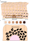
Figure 1
Figure 1
Melanin distribution throughout the epidermis and organization within keratinocytes from distinct phototypes. (A) Melanin dispersion in the epidermis is achieved through the formation of epidermal-melanin units. The accumulation of melanin is higher in the basal layers, regardless of the skin phototype (represented by the numbered circles and labeled from I—lower/lighter phototype—to VI—higher/darker phototype). However, in lower phototypes (lighter skins), less melanin is concentrated in the basal layer, whereas in higher phototypes (darker skins), higher levels can be found in the basal layer, and melanin is also present in the immediate suprabasal layers. (B) Melanin organizes differently within keratinocytes derived from different phototypes. In lower phototypes, melanin is mainly stored in clusters of several melanin granules within a single membrane organelle, while higher phototypes prominently accumulate single granules in single membrane organelles.
Figure 2
Figure 2
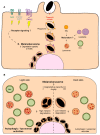
Melanin transfer from melanocytes to keratinocytes. Four different models have been proposed to explain the mechanism(s) of melanin transfer from melanocytes (dark-colored) to keratinocytes (light-colored), including the cytophagocytosis of melanocyte dendrites by keratinocytes; the direct fusion of melanocyte and keratinocyte membranes; the shedding of melanosome-laden globules by melanocytes; and the coupled exocytosis/phagocytosis of the melanin core. Despite the evidence published using different models, recent studies using human and mouse cell lines, as well as more complex models such as reconstructed human skins/epidermis and skin biopsies, have supported the transfer of melanin in globules or as melanocores.
Figure 3
Figure 3
Melanin exocytosis by melanocytes and internalization by keratinocytes. (A) Melanin exocytosis in physiological conditions is thought to occur at the tips of melanocyte dendrites, requiring melanosome transport to the cell periphery. Anterograde long-range transport occurs via microtubules and is mediated by the complex Rab1-SKIP-kinesin 1. On the other hand, retrograde transport is regulated by Rab36-melanoregulin-RILP-DCTN1 and/or Rab44-dynactin-dynein. Melanosome positioning results from the balance between anterograde and retrograde transport. Since retrograde transport overrides anterograde transport, melanosomes tend to accumulate in the perinuclear region. Melanosome peripheral positioning is dependent on the tripartite complex composed of Rab27a-melanophilin-myosin Va. The small GTPase Rab11b and the exocyst complex subunits Exo70 and Sec8 were found to regulate basal melanin exocytosis and transfer. Furthermore, Rab3a enhances melanin exocytosis and transfer to keratinocytes under stimulation by soluble factors derived from differentiated keratinocytes. Whether melanosome-laden globules are also secreted from melanocytes using the same molecular regulators is not known (marked by “?”). (B) Recent studies found key differences in the molecular players involved in melanin recognition and internalization by keratinocytes according to the way melanin is presented to keratinocytes, i.e., “naked” melanocores or membrane-bound melanosomes. Melanocores were shown to be phagocytosed in a PAR-2-dependent manner by keratinocytes, requiring Rac1 and Cdc42 for efficient internalization. The existence and identity of the phagocytic receptor that recognizes melanocores remains elusive (marked by “?”) In contrast, purified membrane-bound melanosomes are internalized through macropinocytosis in a PAR-2-independent manner, requiring RhoA and CtBP1/BARS. Whether the internalization of melanosome-laden globules requires the same regulators as melanosomes remains to be determined (marked by “*”). Additionally, FGFR2b/KGFR and TLR3 stimulation were shown to enhance the internalization of melanosomes and melanocores, although the mechanisms were not characterized. Whether the existence of different internalization routes affects melanokerasome processing is not known (marked by “!”).
Figure 4
Figure 4
Melanin processing within keratinocytes. (A) Recent studies have proposed that transferred melanin inside keratinocytes is surrounded by early and late endocytic markers. Whether this organelle undergoes further maturation is not known, but our recent observations suggest that melanin is stored in a specialized organelle that we named melanokerasome (MKS), which has a poorly acidic lumen and non-degradative properties. The contribution of other organelles to the biogenesis of MKSs is not fully understood, despite the possible involvement of early endocytic, late endocytic, and autophagic vesicles. Additionally, the relevance of receptor signaling (e.g., PAR-2, KGFR/FGFR2, and TLR3 or phagocytic receptors) for the trafficking and biogenesis of MKSs is not known. (B) Autophagy has been reported to regulate melanin degradation within keratinocytes, although the mechanisms are not understood. Moreover, autophagic and lysosomal activities are higher in lighter skin when compared to darker skin. Nevertheless, it is not fully understood whether MKSs from distinct phototypes present different properties regarding their ability to allow melanin persistence or degradation (marked by arrows–more arrows mean lower degradative capacity and acidity), nor the contribution of autophagic/lysosomal activities (marked by “?”) for this. EEs—Early endosomes; LEs—Late endosomes; MVBs—Multivesicular bodies; AVs—Autophagic vesicles; Lys—lysosomes.
Figure 5
Figure 5
Melanin trafficking and positioning within keratinocytes. The accumulation of melanin in the supranuclear region of keratinocytes is crucial for its photoprotective properties in the skin. Despite the importance of this process, the molecular mechanisms involved are still poorly understood. Melanin is known to be retrogradely transported along microtubules in a process mediated by the dynein-dynactin motor complex. It is not known if an adaptor protein (or proteins, marked by “?”) is involved in this process or how the transport is regulated.

References

    1. Grice E.A., Segre J.A. The skin microbiome. Nat. Rev. Microbiol. 2011;9:244–253. doi: 10.1038/nrmicro2537. - DOI - PMC - PubMed
    1. Solanas G., Benitah S.A. Regenerating the skin: A task for the heterogeneous stem cell pool and surrounding niche. Nat. Rev. Mol. Cell Biol. 2013;14:737–748. doi: 10.1038/nrm3675. - DOI - PubMed
    1. Kolarsick P.A.J., Kolarsick M.A., Goodwin C. Anatomy and Physiology of the Skin. J. Dermatol. Nurses Assoc. 2011;3:203–213. doi: 10.1097/JDN.0b013e3182274a98. - DOI
    1. Plonka P.M., Passeron T., Brenner M., Tobin D.J., Shibahara S., Thomas A., Slominski A., Kadekaro A.L., Hershkovitz D., Peters E., et al. What are melanocytes really doing all day long…? Exp. Dermatol. 2009;18:799–819. doi: 10.1111/j.1600-0625.2009.00912.x. - DOI - PMC - PubMed
    1. Cox N.H. Fitzpatrick’s Dermatology in General Medicine. Br. J. Dermatol. 2004;150:794. doi: 10.1111/j.1365-2133.2004.6032_1.x. - DOI

LinkOut - more resources