. 2023 Jul 11;24(14):11329.
doi: 10.3390/ijms241411329.

Suppression of Indoxyl Sulfate Accumulation Reduces Renal Fibrosis in Sulfotransferase 1a1-Deficient Mice

Affiliations

Suppression of Indoxyl Sulfate Accumulation Reduces Renal Fibrosis in Sulfotransferase 1a1-Deficient Mice

Huixian Hou et al. Int J Mol Sci. .

Abstract

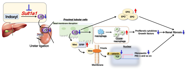
Renal fibrosis is the final manifestation of chronic kidney disease (CKD); its prevention is vital for controlling CKD progression. Indoxyl sulfate (IS), a typical sulfate-conjugated uremic solute, is produced in the liver via the enzyme sulfotransferase (SULT) 1A1 and accumulates significantly during CKD. We investigated the toxicopathological role of IS in renal fibrosis using Sult1a1-KO mice and the underlying mechanisms. The unilateral ureteral obstruction (UUO) model was created; kidney IS concentrations, inflammation, and renal fibrosis were assessed on day 14. After UUO treatment, inflammation and renal fibrosis were exacerbated in WT mice, with an accumulation of IS in the kidney. However, they were significantly suppressed in Sult1a1-KO mice. CD206+ expression was upregulated, and β-catenin expression was downregulated in Sult1a1-KO mice. To confirm the impact of erythropoietin (EPO) on renal fibrosis, we evaluated the time-dependent expression of EPO. In Sult1a1-KO mice, EPO mRNA expression was improved considerably; UUO-induced renal fibrosis was further attenuated by recombinant human erythropoietin (rhEPO). Thus, UUO-induced renal fibrosis was alleviated in Sult1a1-KO mice with a decreased accumulation of IS. Our findings confirmed the pathological role of IS in renal fibrosis and identified SULT1A1 as a new therapeutic target enzyme for preventing and attenuating renal fibrosis.

Keywords: indoxyl sulfate; renal fibrosis; sulfotransferase 1a1-deficient mice.


Conflict of interest statement

The authors declare no conflict of interest.

Figures

Figure 1
Figure 1
Effect of Sult1a1-KO on IS concentration in serum and kidney. (A) Serum BUN concentration in WT and Sult1a1-KO mice. (B) Sult1a1 gene expression checked using RT-PCR. (CE) IS concentration in serum, kidney, and liver calculated via LC-MS/MS. Each value represents the mean ± SD of 3–9 mice. ** p < 0.01, ns: not significant.
Figure 2
Figure 2
Effect of Sult1a1-KO on inflammation, renal fibrosis. (A,B) gene expression of renal fibrosis markers (col1a1, fibronectin) checked using RT-PCR. (CE) gene expression of inflammatory cytokines (TNF-α, IL-1β, IL-6) checked using RT-PCR. Each value represents the mean ± SD of 3–9 mice. * p < 0.05, ** p < 0.01, ns: not significant.
Figure 3
Figure 3
Effect of Sult1a1-KO on UUO-induced renal fibrosis. (A) collagen deposition (the red part) in the kidney confirmed by Sirius red staining. The scale bar = 50 μm. (B) Western blot experiment confirmed the expression of α-SMA. Each value represents the mean ± SD of 3–9 mice. * p < 0.05, ** p < 0.01.
Figure 4
Figure 4
Effect of Sult1a1-KO on UUO-induced CD206+ macrophage infiltration. (A,B) gene expression of F4/80 and CD206+ checked using RT-PCR. (C) immunostaining of CD206. The scale bar = 50 μm. (D) apoptotic cells checked via TUNEL staining. The scale bar = 50 μm. Each value represents the mean ± SD of 7–8 mice. * p < 0.05, ** p < 0.01, ns: not significant.
Figure 5
Figure 5
Effect of Sult1a1-KO on Wnt/β-catenin signaling activation. (A) gene expression of Wnt4 checked using RT-PCR. (B) gene expression of Sfrp5 checked using RT-PCR. (C) gene expression of β-catenin checked using RT-PCR. (D) protein expression of β-catenin checked via western blot. Each value represents the mean ± SD of 7–8 mice. ** p < 0.01, ns: not significant.
Figure 6
Figure 6
Effect of rhEPO treatment and Sult1a1-KO on renal fibrosis. (A) time-dependent expression of EPO gene expression tested using RT-PCR. (B) Protein expression of α-SMA checked using western blot. (C) Collagen deposition (the red part) in the kidney confirmed via Sirius red staining. The scale bar = 50 μm. Each value represents the mean ± SD of 3–8 mice. * p < 0.05, ** p < 0.01.
Figure 7
Figure 7
Summary of the mechanisms underlying UUO-induced renal fibrosis suppressed in Sult1a1-KO mice. After the unilateral ureter was obstructed in Sult1a1-KO mice, IS accumulation was diminished in the serum and renal tissues due to a lack of Sult1a1 activity. UUO-induced renal fibrosis was suppressed, accompanied by decreased IS concentration. Furthermore, Sult1a1 inhibition induced the infiltration of CD206+ macrophages, suppressing ongoing inflammation and, as a result, renal fibrosis. Inactivated Wnt/β-catenin signaling and increased EPO production also help to reduce renal fibrosis.

Similar articles

Cited by

References

    1. Zhang L., Zhang P., Wang F., Zuo L., Zhou Y., Shi Y., Li G., Jiao S., Liu Z., Liang W., et al. Prevalence and factors associated with CKD: A population study from Beijing. Am. J. Kidney Dis. 2008;51:373–384. doi: 10.1053/j.ajkd.2007.11.009. - DOI - PubMed
    1. Sharma S.K., Zou H., Togtokh A., Ene-Iordache B., Carminati S., Remuzzi A., Wiebe N., Ayyalasomayajula B., Perico N., Remuzzi G., et al. Burden of CKD, proteinuria, and cardiovascular risk among Chinese, Mongolian, and Nepalese participants in the International Society of Nephrology screening programs. Am. J. Kidney Dis. 2010;56:915–927. doi: 10.1053/j.ajkd.2010.06.022. - DOI - PubMed
    1. Lv J.C., Zhang L.X. Prevalence and Disease Burden of Chronic Kidney Disease. Adv. Exp. Med. Biol. 2019;1165:3–15. doi: 10.1007/978-981-13-8871-2_1. - DOI - PubMed
    1. Liu Y. Cellular and molecular mechanisms of renal fibrosis. Nat. Rev. Nephrol. 2011;7:684–696. doi: 10.1038/nrneph.2011.149. - DOI - PMC - PubMed
    1. Miyazaki T., Ise M., Hirata M., Endo K., Ito Y., Seo H., Niwa T. Indoxyl sulfate stimulates renal synthesis of transforming growth factor-beta 1 and progression of renal failure. Kidney Int. Suppl. 1997;63:S211–S214. - PubMed