. 2023 Jul 14;24(14):11473.
doi: 10.3390/ijms241411473.

Triplin: Mechanistic Basis for Voltage Gating

Affiliations

Triplin: Mechanistic Basis for Voltage Gating

Marco Colombini et al. Int J Mol Sci. .

Abstract

The outer membrane of Gram-negative bacteria contains a variety of pore-forming structures collectively referred to as porins. Some of these are voltage dependent, but weakly so, closing at high voltages. Triplin, a novel bacterial pore-former, is a three-pore structure, highly voltage dependent, with a complex gating process. The three pores close sequentially: pore 1 at positive potentials, 2 at negative and 3 at positive. A positive domain containing 14 positive charges (the voltage sensor) translocates through the membrane during the closing process, and the translocation is proposed to take place by the domain entering the pore and thus blocking it, resulting in the closed conformation. This mechanism of pore closure is supported by kinetic measurements that show that in the closing process the voltage sensor travels through most of the transmembrane voltage before reaching the energy barrier. Voltage-dependent blockage of the pores by polyarginine, but not by a 500-fold higher concentrations of polylysine, is consistent with the model of pore closure, with the sensor consisting mainly of arginine residues, and with the presence, in each pore, of a complementary surface that serves as a binding site for the sensor.

Keywords: cooperativity; kinetics; polyarginine; pore; porin; prokaryote; voltage dependence; voltage sensor.


Conflict of interest statement

The authors declare no conflict of interest.

Figures

Figure 1
Figure 1
Voltage gating in a single Triplin. On the left side, a triangular voltage wave (+75/−77mV; 30 mHz) results in no pore closure. In region “A”, a +69 mV potential was applied to the cis compartment. At point “B”, pore 1 closed. The resumption of the triangular wave caused gating of pores 2 and 3. “C” indicates the location of pore 2 closure and “D” the locations of pore 2 reopening. “E” is the point at which pore 3 closed and at “F” pores 2 and 3 opened simultaneously.
Figure 2
Figure 2
Model of the gating of Triplin. The top of the structure is the cis side of the membrane, the side from which Triplin is inserted. The bottom of the structure is the trans side and that is the side maintained at virtual ground by the amplifier. The numbers refer to pores 1, 2 and 3. All indicated voltages refer to the cis side. For simplicity, the closed state of the pore is illustrated as a result of blockage by a single loop of the beta barrel, but, of course, multiple loops may be involved. Blue regions are positively charged whereas red are negatively charged. From Figure 14 in ref [1].
Figure 3
Figure 3
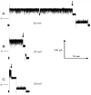
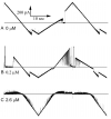
Pore 2 closing time at three different voltages, labeled (AC). In all records, the initial voltage was 10 mV (short segment on left). The voltage was then switched to the indicated value, and at the point indicated by the arrow, pore 2 closed. The voltage was then switched to 10 mV, and shortly thereafter, the pore reopened. Zero current is indicated by the short line on the left side. The inset shows a plot of the average closure time (the time constant) as a function of voltage for that experiment.
Figure 4
Figure 4
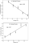
Voltage dependence of the opening and closing time constants for pore 2. The error bars are standard errors of the mean of 20 measurements.
Figure 5
Figure 5
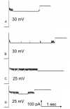
Pore 3 closure interferes with the opening of pore 2. Four sample records are illustrated, two taken at 25 mV (C,D) and two at 30 mV (A,B). Pore 2 was closed at −40 mV, and then the positive voltage indicated was applied. The downward events are pore 3 closures: some are transient and other are long-lived. In the case of record (C), the reopening of pore 3 after its closure in the middle of the record, took place at a time beyond the end of the record shown. Hence, pore 2 opening is not visible in the record shown.
Figure 6
Figure 6
Pore 3 closure blocks pore 2 opening. Pore 2 opening at various applied voltages was measured either by ignoring pore 3 closure (circle and square symbols) or by subtracting the time during which pore 3 was closed (triangles). Error bars are SEM of 18 to 22 measurements.
Figure 7
Figure 7
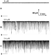
Transient blocking events by the addition of the indicated amount of polyarginine to the cis side of a membrane containing a single Triplin with all pores open. The three records shown (AC) are typical samples of the recorded blockage events. The zero current level is indicated just below the record. All records were collected in the presence of a 40 mV applied potential.
Figure 8
Figure 8
Long-lived pore blockage in the presence of 0.4 µg/ml polyarginine. A single Triplin was still gating as demonstrated by the triangular voltage wave (+72/−71 mV) on the far left side. Pore 2 closure was followed by pore 3 closure and then simultaneous pore 2 and 3 opening at high positive voltages allowing polyarginine to block transiently. This was followed by applying a constant voltage (+50 mV) resulting in both transient and long-lived blockages. The expanded regions show that often both pores were blocked simultaneously but at times unblocking took place in two separate events. These regions were only expanded in the time axis.
Figure 9
Figure 9
Voltage-dependent block of Triplin by polyarginine. A triangular voltage wave (30 mHz; +72 to −71 mV) was applied to a membrane containing a single Triplin activated by closing pore 1. The horizontal lines are the zero current levels. Polyarginine was added sequentially, and sample records are illustrated. Record (A) was taken before polyarginine addition, followed later by (B) and much later by (C). The amount of polyarginine present in the cis compartment during each recording is indicated.
Figure 10
Figure 10
Voltage dependence of the formation of long-lived pore blocks by polyaginine at the indicated concentration. The power dependence of the exponential fit to the data beginning at 30 mV is indicated next to each curve.
Figure 11
Figure 11
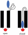
Model of the gating process used by each pore. Left is the open state with the sensor (blue) out of the pore. The red negative domain is close to the other end of the pore and proposed to be responsible for the selectivity of the pore and the rectification. Right shows the obstructed pore with the sensor (blue) interacting with a negative domain (red).

References

    1. Colombini M., Barnes K., Chang K.-T., Younis M.H., Aguilella V.M. Triplin: Functional probing of its structure and the dynamics of the voltage-gating process. Int. J. Mol. Sci. 2022;23:13765. doi: 10.3390/ijms232213765. - DOI - PMC - PubMed
    1. Schindler H., Rosenbusch J.P. Matrix protein in planar membranes: Clusters of channels in a native environment and their functional reassembly. Proc. Natl. Acad. Sci. USA. 1981;78:2302–2306. doi: 10.1073/pnas.78.4.2302. - DOI - PMC - PubMed
    1. Van Gelder P., Saint N., Phale P., Eppens E.F., Prilipov A., Van Boxtel R., Rosenbusch J.P., Tommassen J. Voltage sensing in the PhoE and OmpF outer membrane porins of Escherichia coli: Role of charged residues. J. Mol. Biol. 1997;269:468–472. doi: 10.1006/jmbi.1997.1063. - DOI - PubMed
    1. Robertson K.M., Tieleman D.P. Molecular basis of voltage gating of OmpF porin. Biochem. Cell Biol. 2002;80:517–523. doi: 10.1139/o02-145. - DOI - PubMed
    1. Nikaido H. Molecular basis of bacterial outer membrane permeability revisited. Microbiol. Mol. Biol. Rev. 2003;67:593–656. doi: 10.1128/MMBR.67.4.593-656.2003. - DOI - PMC - PubMed

LinkOut - more resources