. 2023 Jul 17;24(14):11551.
doi: 10.3390/ijms241411551.

Effects of Probiotics on Colitis-Induced Exacerbation of Alzheimer's Disease in AppNL-G-F Mice

Affiliations

Effects of Probiotics on Colitis-Induced Exacerbation of Alzheimer's Disease in AppNL-G-F Mice

Bijayani Sahu et al. Int J Mol Sci. .

Abstract

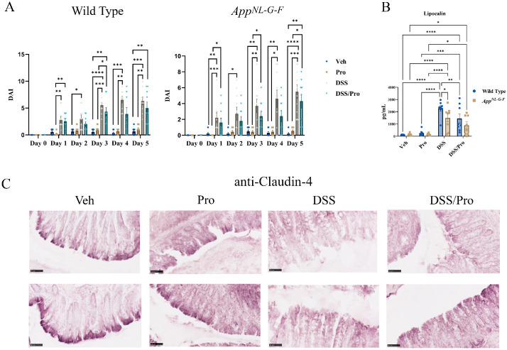
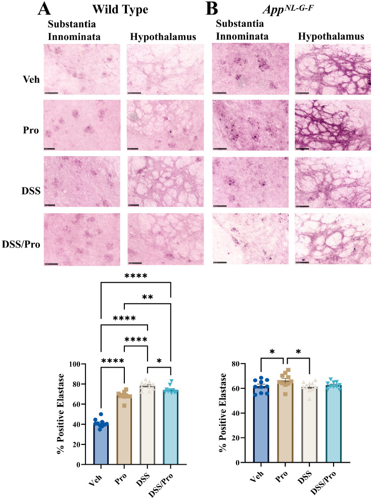
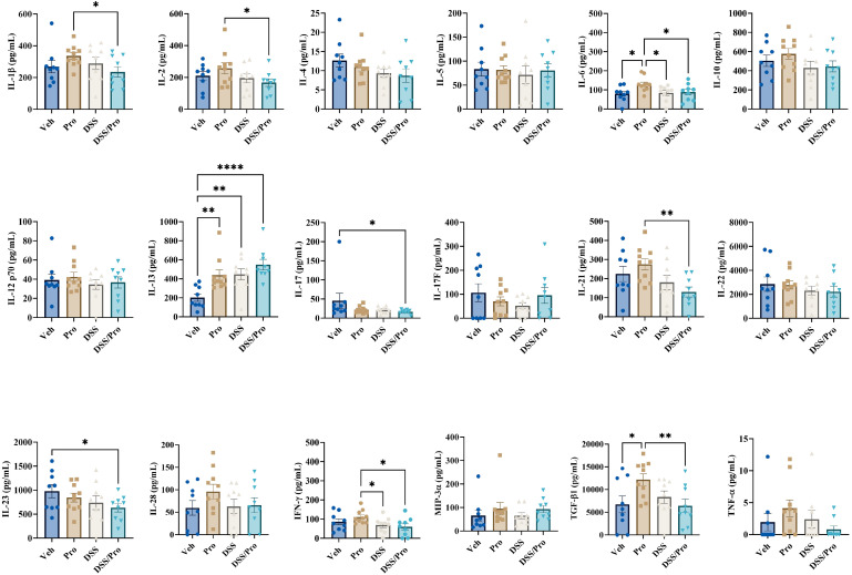
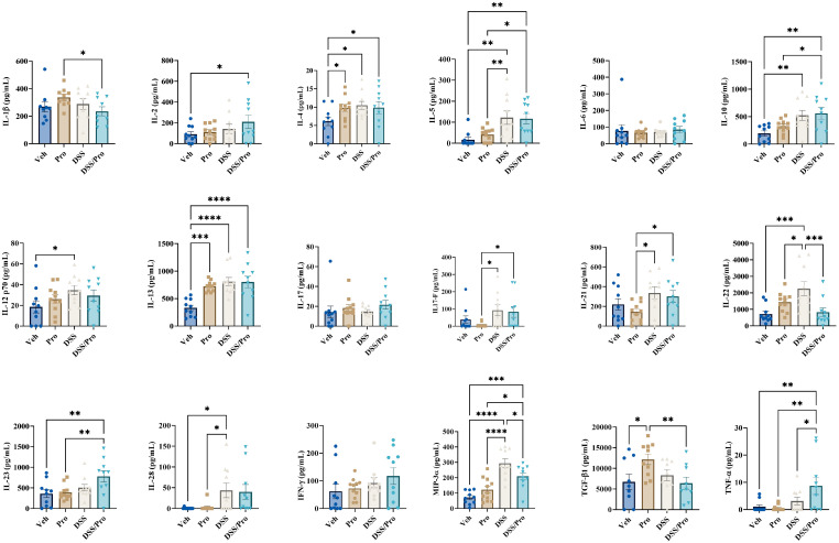
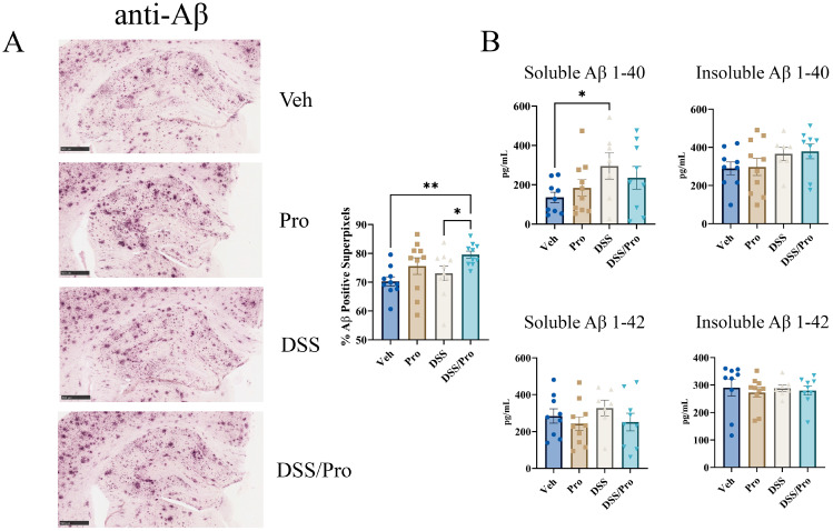
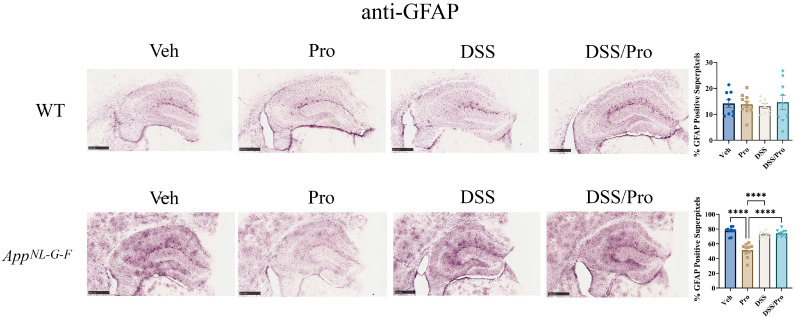
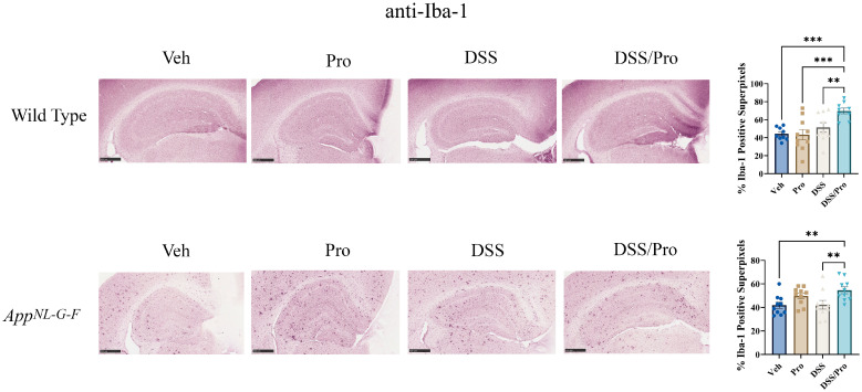
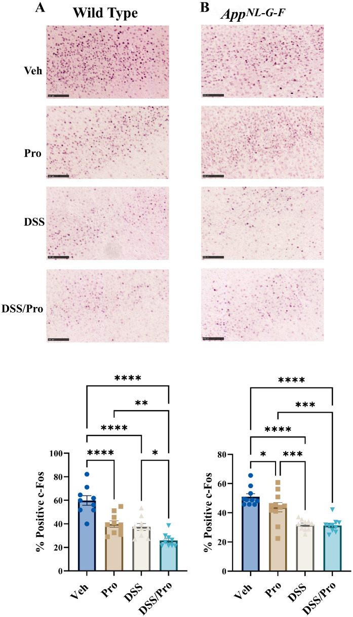
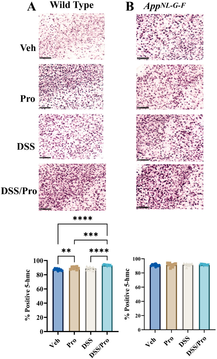
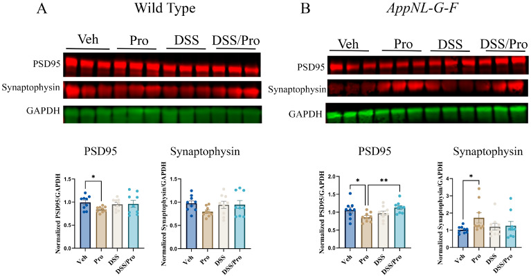
Alzheimer's disease (AD) is characterized by progressive cognitive decline and is a leading cause of death in the United States. Neuroinflammation has been implicated in the progression of AD, and several recent studies suggest that peripheral immune dysfunction may influence the disease. Continuing evidence indicates that intestinal dysbiosis is an attribute of AD, and inflammatory bowel disease (IBD) has been shown to aggravate cognitive impairment. Previously, we separately demonstrated that an IBD-like condition exacerbates AD-related changes in the brains of the AppNL-G-F mouse model of AD, while probiotic intervention has an attenuating effect. In this study, we investigated the combination of a dietary probiotic and an IBD-like condition for effects on the brains of mice. Male C57BL/6 wild type (WT) and AppNL-G-F mice were randomly divided into four groups: vehicle control, oral probiotic, dextran sulfate sodium (DSS), and DSS given with probiotics. As anticipated, probiotic treatment attenuated the DSS-induced colitis disease activity index in WT and AppNL-G-F mice. Although probiotic feeding significantly attenuated the DSS-mediated increase in WT colonic lipocalin levels, it was less protective in the AppNL-G-F DSS-treated group. In parallel with the intestinal changes, combined probiotic and DSS treatment increased microglial, neutrophil elastase, and 5hmC immunoreactivity while decreasing c-Fos staining compared to DSS treatment alone in the brains of WT mice. Although less abundant, probiotic combined with DSS treatment demonstrated a few similar changes in AppNL-G-F brains with increased microglial and decreased c-Fos immunoreactivity in addition to a slight increase in Aβ plaque staining. Both probiotic and DSS treatment also altered the levels of several cytokines in WT and AppNL-G-F brains, with a unique increase in the levels of TNFα and IL-2 being observed in only AppNL-G-F mice following combined DSS and probiotic treatment. Our data indicate that, while dietary probiotic intervention provides protection against the colitis-like condition, it also influences numerous glial, cytokine, and neuronal changes in the brain that may regulate brain function and the progression of AD.

Keywords: Alzheimer’s; amyloid; colitis; intestine.


Conflict of interest statement

The authors declare no conflict of interest.

Figures

Figure 1
Figure 1
Probiotic treatment had minimal effects on DSS-induced colitis disease activity index (DAI), lipocalin levels, and claudin-4 immunoreactivity in C57BL/6J wild type (WT) and AppNL-G-F mice. Male C57BL/6J wild type and AppNL-G-F mice were given diluted MediGel in water (1:1) ad libitum or the probiotic resuspended in diluted MediGel starting at week 0 and until week 8. At weeks 3 and 5, DSS was provided in the diluted MediGel (2% final concentration) for three days per cycle. Mice were allowed to recover until week 8 with or without maintained exposure to the probiotic. (A) A colitis-like disease (DAI) was assessed in vehicle, probiotic (Pro), DSS, and DSS/Pro treatment groups in male wild type and AppNL-G-F mice. The DAI was monitored on the second 3-day cycle of 2% DSS exposure and 2 days afterwards (day 0–day 5) in all treatment groups. (B) Upon completion of the probiotic feeding paradigm at 8 weeks, the wild type and AppNL-G-F mice were collected before colon lipocalin levels were quantified by ELISA. (C) Claudin 4 immunoreactivity was examined in colons of both wild type and AppNL-G-F colons using Vector VIP as the chromogen. Representative images are shown (scale bar 50 μm). Non-parametric one-way ANOVA (Kruskal–Wallis test) followed by Dunn’s multiple comparisons test was used to determine statistical differences. Results are presented as mean ± SEM, * p < 0.05, ** p < 0.01, *** p < 0.001, and **** p < 0.0001.
Figure 2
Figure 2
DSS and probiotic increased brain neutrophil elastase immunoreactivity in wild type but not AppNL-G-F mice. After the 8 weeks of probiotic feeding, brains of (A) wild type and (B) AppNL-G-F mice were fixed and serial sectioned (40 µm) for anti-neutrophil elastase immunohistochemistry. Representative neutrophil elastase immunohistochemical staining images (20×) of the substantia innominata and hypothalamus are shown (scale bar: 100 µm). Elastase positive cell counts were quantified from an entire hemibrain coronal section from 3 to 10 sections per brain in each group. The positive counts were then measured as a percentage of the annotated area. One-way ANOVA followed by uncorrected Fisher’s LSD test was used to determine statistical differences. Results are presented as mean values + SEM, * p < 0.05, ** p < 0.01, and **** p < 0.0001.
Figure 3
Figure 3
DSS and probiotic altered numerous cytokine levels in the cortices of wild type mice. After completing the probiotic feeding paradigm at 8 weeks, C57BL/6J wild type mice were collected, and temporal cortex cytokine levels were quantified by commercial slide array. One-way ANOVA followed by uncorrected Fisher’s LSD test was used to determine statistical differences. Results are presented as mean ± SEM, * p < 0.05, ** p < 0.01, and **** p < 0.0001.
Figure 4
Figure 4
DSS and probiotic altered numerous cytokine levels in the cortices of AppNL-G-F mice. After completing the probiotic feeding paradigm at 8 weeks, AppNL-G-F mice were collected, and temporal cortex cytokine levels were quantified by commercial slide array. One-way ANOVA followed by uncorrected Fisher’s LSD test was used to determine statistical differences. Results are presented as mean ± SEM, * p < 0.05, ** p < 0.01, *** p < 0.001, and **** p < 0.0001.
Figure 5
Figure 5
DSS increased hippocampal Aβ levels in AppNL-G-F mouse brains. After completing the probiotic feeding paradigm at 8 weeks, wild type and AppNL-G-F mice were collected. (A) Left hemispheres of AppNL-G-F mice were fixed and serial sectioned (40 µm) for anti-Aβ immunohistochemistry. Percent Aβ positive super pixels in the hippocampus were determined from three sections per mouse in each condition. One-way ANOVA followed by uncorrected Fisher’s LSD test was used to determine statistical differences. Representative 5× images are shown (scale bar 500 μm). Results are presented as mean ± SEM, * p < 0.05, ** p < 0.01 (n = 10). (B) Hippocampal levels of human-soluble and insoluble Aβ 1-40 and 1-42 were quantified by ELISA from AppNL-G-F lysates. One-way ANOVA followed by uncorrected Fisher’s LSD test was used to determine statistical differences. Results are presented as mean ± SEM, * p < 0.05 and ** p < 0.01.
Figure 6
Figure 6
Probiotic treatment reduced astrogliosis in the hippocampus of AppNL-G-F mice. After 8 weeks of probiotic feeding, the brains of the AppNL-G-F mice and wild type mice were fixed and serial sectioned (40 µm) for anti-GFAP immunohistochemistry. The percentage of GFAP-positive super pixels in the hippocampus were determined from three sections per mouse in each condition. One-way ANOVA followed by uncorrected Fisher’s LSD test was used to determine statistical differences. Results are presented as mean ± SEM; **** p < 0.0001. Representative 5X images are shown (scale bar: 500 μm).
Figure 7
Figure 7
Combined DSS and probiotic treatment increased microgliosis in the hippocampus of wild type and AppNL-G-F mice. After the 8 weeks of probiotic feeding, the brains of AppNL-G-F and wild type mice were fixed and serial sectioned (40 µm) for anti-Iba-1 immunohistochemistry. The percentage of Iba-1-positive super pixels in the hippocampus were determined from three sections per mouse in each condition. One-way ANOVA followed by uncorrected Fisher’s LSD test was used to determine statistical differences. Results are presented as mean ± SEM, ** p < 0.01, and *** p < 0.001. Representative 5× images are shown (scale bar: 500 μm).
Figure 8
Figure 8
DSS and probiotic attenuated c-Fos staining in the brains of wild type and AppNL-G-F mice. After 8 weeks of probiotic feeding, brains of (A) wild type and (B) AppNL-G-F mice were fixed and serial sectioned (40 µm) for anti-c-Fos immunohistochemistry. Representative images (20×) from the parietal cortex are shown (scale bar: 100 μm). c-Fos-positive cell counts were quantified from an entire hemibrain coronal section from 3 to 10 sections per brain in each group. The positive counts were then measured as a percentage of the annotated area. Values are presented as mean ± SEM. One-way ANOVA followed by uncorrected Fisher’s LSD test was used to determine statistical differences; * p < 0.5, ** p < 0.01, *** p < 0.001, and **** p < 0.0001.
Figure 9
Figure 9
Probiotic and DSS increased 5hmC staining only in the brains of wild type mice. After 8 weeks of probiotic feeding, brains of (A) wild type and (B) AppNL-G-F mice were fixed and serial sectioned (40 µm) for anti-5hmC immunohistochemistry. Representative images (20×) from the parietal cortex are shown (scale bar 100 μm). 5hmC positive cell counts were quantified from an entire hemibrain coronal section from 3 to 10 sections per brain in each group. The positive counts were then measured as a percentage of the annotated area. Values are presented as mean ± SEM. One-way ANOVA followed by uncorrected Fisher’s LSD test was used to determine statistical differences; ** p < 0.01, *** p < 0.001, and **** p < 0.0001.
Figure 10
Figure 10
DSS and probiotic effects on presynaptic or postsynaptic protein levels in the brains of wild type and AppNL-G-F mice. After 8 weeks of probiotic feeding, the temporal cortices of (A) wild type and (B) AppNL-G-F mice treated with or without DSS were lysed, and the proteins were resolved by SDS-PAGE for Western blot analysis using antibodies against PSD95, synaptophysin, and GAPDH (loading control). Data from Western blots are graphed as mean ± SEM of PSD95 or synaptophysin values normalized to their respective GAPDH. One-way ANOVA followed by uncorrected Fisher’s LSD test was used to determine statistical differences; * p < 0.05 and ** p < 0.001.

Similar articles

Cited by

References

    1. Bondi M.W., Edmonds E.C., Salmon D.P. Alzheimer’s Disease: Past, Present, and Future. J. Int. Neuropsychol. Soc. 2017;23:818–831. doi: 10.1017/S135561771700100X. - DOI - PMC - PubMed
    1. Kumar A., Sidhu J., Goyal A., Tsao J.W. Alzheimer Disease. StatPearls; Treasure Island, FL, USA: 2022.
    1. Tiwari S., Atluri V., Kaushik A., Yndart A., Nair M. Alzheimer’s disease: Pathogenesis, diagnostics, and therapeutics. Int. J. Nanomed. 2019;14:5541–5554. doi: 10.2147/IJN.S200490. - DOI - PMC - PubMed
    1. Collaborators G.B.D.D.F. Estimation of the global prevalence of dementia in 2019 and forecasted prevalence in 2050: An analysis for the Global Burden of Disease Study 2019. Lancet Public Health. 2022;7:e105–e125. doi: 10.1016/S2468-2667(21)00249-8. - DOI - PMC - PubMed
    1. Huang L., Lu Z., Zhang H., Wen H., Li Z., Liu Q., Wang R. A Novel Strategy for Alzheimer’s Disease Based on the Regulatory Effect of Amyloid-β on Gut Flora. J. Alzheimer’s Dis. JAD. 2022:1–13. doi: 10.3233/JAD-220651. - DOI - PMC - PubMed