. 2023 Jul 19;24(14):11641.
doi: 10.3390/ijms241411641.

Epigenetic Modulation of Inflammatory Pathways in Myometrial Stem Cells and Risk of Uterine Fibroids

Affiliations

Epigenetic Modulation of Inflammatory Pathways in Myometrial Stem Cells and Risk of Uterine Fibroids

Qiwei Yang et al. Int J Mol Sci. .

Abstract

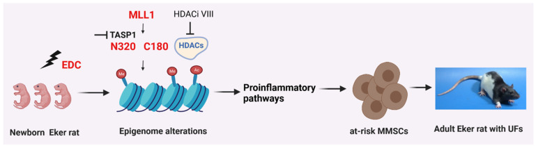
The period during which tissue and organ development occurs is particularly vulnerable to the influence of environmental exposures. However, the specific mechanisms through which biological pathways are disrupted in response to developmental insults, consequently elevating the risk of hormone-dependent diseases, such as uterine fibroids (UFs), remain poorly understood. Here, we show that developmental exposure to the endocrine-disrupting chemical (EDC), diethylstilbestrol (DES), activates the inflammatory pathways in myometrial stem cells (MMSCs), which are the origin of UFs. Significantly, the secretome of reprogrammed MMSCs enhances the expression of critical inflammation-related genes in differentiated myometrial cells through the paracrine mechanism, which amplifies pro-inflammatory and immune suppression signaling in the myometrium. The expression of reprogrammed inflammatory responsive genes (IRGs) is driven by activated mixed-lineage leukemia protein-1 (MLL1) in MMSCs. The deactivation of MLL reverses the reprogramming of IRG expression. In addition, the inhibition of histone deacetylases (HDACs) also reversed the reprogrammed IRG expression induced by EDC exposure. This work identifies the epigenetic mechanisms of MLL1/HDAC-mediated MMSC reprogramming, and EDC exposure epigenetically targets MMSCs and imparts an IRG expression pattern, which may result in a "hyper-inflammatory phenotype" and an increased hormone-dependent risk of UFs later in life.

Keywords: developmental reprogramming; diethylstilbestrol; inflammatory responsive genes; myometrial stem cells; uterine fibroids.


Conflict of interest statement

None of the authors have a financial relationship with a commercial entity that has an interest in the subject of this manuscript.

Figures

Figure 1
Figure 1
Developmental DES-exposure-activated inflammation pathway in MMSCs. (A) Experimental paradigm. Eker rat pups were exposed to VEH and DES at postnatal days 10–12, respectively. The pups were euthanized at five months of age, representing the early adult stage. Myometrial tissues were isolated from the animals and subjected to MMSC isolation using Stro-1/CD44 surface markers. Myometria from five animals were pooled for each treatment. Multi-omics analyses, including RNA-seq and ChIP-seq, were performed to determine the transcriptome and histone modification alterations, respectively. (B) Hallmark gene set analysis between DES-MMSCs and VEH-MMSCs. Inflammation-related pathways are highlighted in red. (C) Upstream regulator and predicted activation analysis via Ingenuity Pathway Analysis (IPA). (D) Pie chart showing the percentage of IRGs that exhibited changes in RNA expression between DES-MMSCs and VEH-MMSCs, as measured via RNA-seq; the cutoff value is 2-fold with an FDR < 0.05. (E) List the top 18 IRGs showing differential upregulation in DES- vs. VEH-MMSCs. (F) List the top 18 IRGs showing downregulation in DES- vs. VEH-MMSCs.
Figure 2
Figure 2
The correlation between RNA expression and H3K4me3-reprogrammed genes. DES-MMSCs and VEH-MMSCs were isolated and subjected to immunoprecipitation with anti-H3K4me3 antibody. ChIP-seq and bioinformatic analysis were performed as described in the Materials and Methods Section. (A) DES-regulated IRGs with enrichment or reduction of H3K4me3. (B) The list of IRGs with H3K4me3 status. Up-DEGs were highlighted in red. Down-DEGs were highlighted in light blue color. Up-H3K4me3 were highlighted in green color. (C) Histograms created using Integrative Genomics Viewer showing H3K4me3 occupancy at Ccl7, Ccl2, Ereg, Cd40, Pcdh7, and Ptger2. For each gene, the upper and lower browser images display an expanded view of a selected region of the H3K4me3 peak distributions in VEH-MMSCs (blue track) and DES-MMSCs (red track). (D) The list of top 20 reprogrammed IRGs showing their cellular functions.
Figure 3
Figure 3
The effect of the secretome of reprogrammed DES-MMSCs on DMC. Serum-free conditioned medium (CM) was prepared from DES-MMSCs and VEH-MMSCs. DMCs from adult rat myometria were grown in the CM from DES-MMSCs and VEH-MMSCs for two days. Bar plots show the relative normalized gene expression of critical inflammation-related genes, including Ccl2, Ccl7, Pdpn, and Lpar1 (left panel), as well as IL-1a, IL-1b, IL-6, IL17, and TNFa (right panel). * p < 0.05; ** p < 0.01, *** p < 0.001, NS, no significant difference.
Figure 4
Figure 4
Specific reprogramming of IRGs in MMSCs. (A) Bar plots showing the differential expression of IRGs, including Itgb3, Kcna3, Tlr2, Sgms2, Pik3R5, and Kif1b, between DES-MMSCs and DES-DMCs. (B) Bar plots showing the differential expression of IRGs, including Aplnr, Lrf7, Itga5, Tnfsf9, and Rela, between DES-MMSCs and DES-DMCs. (C) The correlation between H3K4me3 status and the expression of reprogrammed IRGs (the expression comparison between DES-MMSCs and DES-DMCs, or expression comparison between DES-MMSCs vs VEH-MMSCs). ns: no significant difference. The genes with a grey color background are H3K4me3-enriched genes. The genes with the reduced H3K4me3 are presented with a light blue background. The p-value shows the significant difference in gene expression between Stro-1/CD44 double-positive and double-negative cells. Additionally, the p-values with the blue color show two comparisons of gene expression (Stro-1/CD44 double-positive vs. double-negative cells, or Stro-1+/CD44+ DES vs. VEH) going in the same direction. The p-values with the red color indicated that two comparisons go in the opposite direction, or one comparison shows either up- or down-regulated, and the other shows no significant changes at all. * p < 0.05; ** p < 0.01, *** p < 0.001, **** p < 0.0001. ns: no significant difference. no: no significant changes.
Figure 5
Figure 5
TASP1 is required for the regulation of IRGs expression. (A) Bar plots showing the relative normalized gene expression of Tasp1 RNA levels in DES-MMSCs infected with Tasp1 rat shRNA lentiviral particles (KD) or scrambled particles (Scr). (B) Bar plots showing the relative normalized gene expression of IRGs (Ccl2, Ccl7, Lpar1, and Pdpn) after knockdown of Tasp1. Scr: scramble; KD: knockdown; * p < 0.05; *** p < 0.001, NS: no significant difference.
Figure 6
Figure 6
Inhibition of HDAC activity on the expression of reprogrammed IRGs. Bar plots showing the relative normalized gene expressions of (A) Ccl2, (B) Ccl7, (C) Lpar1, and (D) Pdpn in DES-MMSCs treated with HDAC inhibitor HDACi VIII at concentrations of 2.5 and 5 mg/mL for 1 and 2 days, respectively. * p < 0.05; ** p < 0.01; *** p < 0.001; NS: no significant difference.
Figure 7
Figure 7
Proposed animal model. Exposure to endocrine-disrupting chemicals (EDCs) during development alters the characteristics of myometrial stem cells (MMSCs) and triggers inflammatory pathways within these cells, which are the source of uterine fibroids (UFs). The abnormal expression of reprogrammed inflammatory responsive genes (IRGs) in MMSCs is driven by the activation of mixed-lineage leukemia protein-1 (MLL1). The EDC, acting as an environmental risk factor, disrupts the epigenetic regulation of MMSCs through modifications to histones, leading to the activation of inflammatory pathways, ultimately contributing to the development of UFs. By inhibiting MLL1 and histone deacetylases (HDACs), the reprogramming of IRGs induced by EDC exposure can be reversed. This figure was created using the BioRender software (BioRender.com).

Similar articles

Cited by

References

    1. Yang Q., Ciebiera M., Bariani M.V., Ali M., Elkafas H., Boyer T.G., Al-Hendy A. Comprehensive Review of Uterine Fibroids: Developmental Origin, Pathogenesis, and Treatment. Endocr. Rev. 2022;43:678–719. doi: 10.1210/endrev/bnab039. - DOI - PMC - PubMed
    1. Bulun S.E. Uterine fibroids. N. Engl. J. Med. 2013;369:1344–1355. doi: 10.1056/NEJMra1209993. - DOI - PubMed
    1. Stewart E.A., Laughlin-Tommaso S.K., Catherino W.H., Lalitkumar S., Gupta D., Vollenhoven B. Uterine fibroids. Nat. Rev. Dis. Primers. 2016;2:16043. doi: 10.1038/nrdp.2016.43. - DOI - PubMed
    1. Segars J.H., Parrott E.C., Nagel J.D., Guo X.C., Gao X., Birnbaum L.S., Pinn V.W., Dixon D. Proceedings from the Third National Institutes of Health International Congress on Advances in Uterine Leiomyoma Research: Comprehensive review, conference summary and future recommendations. Hum. Reprod. Update. 2014;20:309–333. doi: 10.1093/humupd/dmt058. - DOI - PMC - PubMed
    1. Yang Q., Diamond M.P., Al-Hendy A. Early Life Adverse Environmental Exposures Increase the Risk of Uterine Fibroid Development: Role of Epigenetic Regulation. Front. Pharmacol. 2016;7:40. doi: 10.3389/fphar.2016.00040. - DOI - PMC - PubMed