. 2023 Jul 19;24(14):11658.
doi: 10.3390/ijms241411658.

Effective Healing of Staphylococcus aureus-Infected Wounds in Pig Cathelicidin Protegrin-1-Overexpressing Transgenic Mice

Affiliations

Effective Healing of Staphylococcus aureus-Infected Wounds in Pig Cathelicidin Protegrin-1-Overexpressing Transgenic Mice

Nagasundarapandian Soundrarajan et al. Int J Mol Sci. .

Abstract

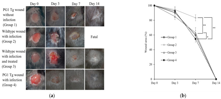
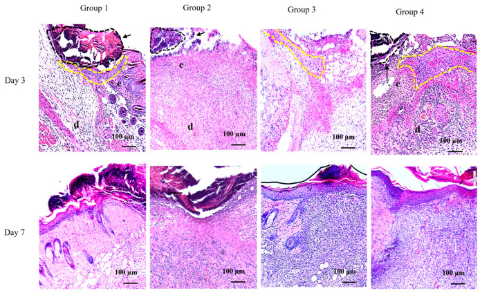
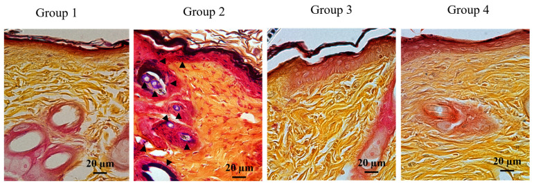
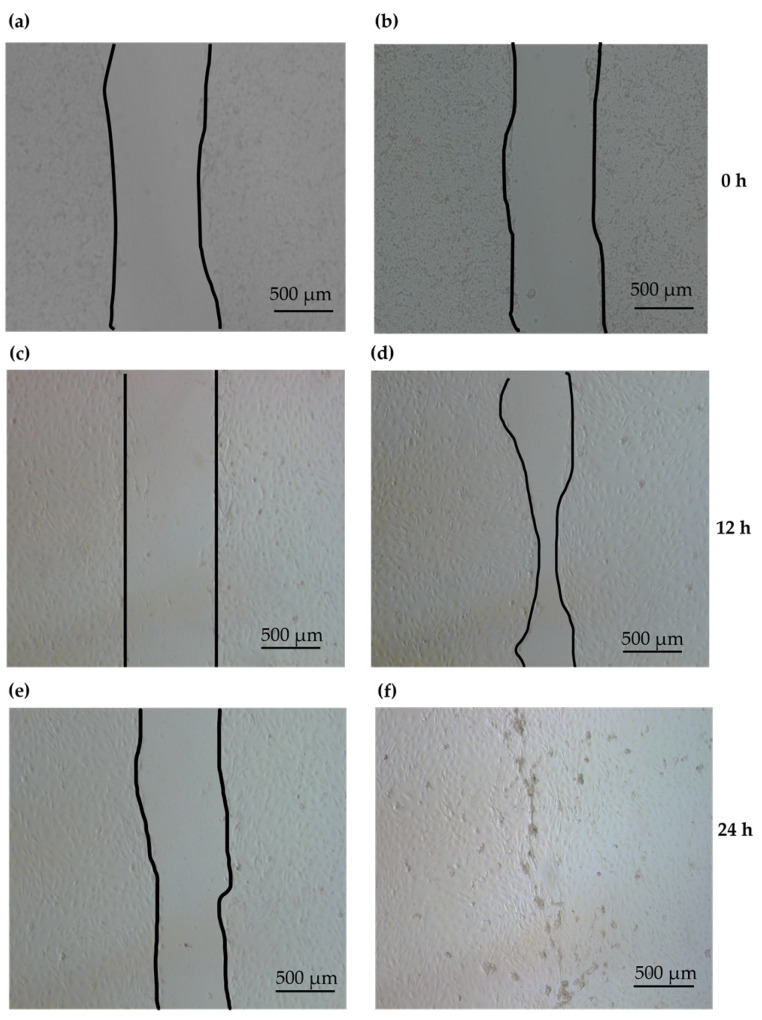
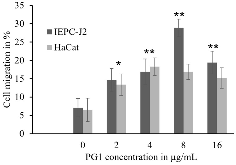
Antimicrobial peptides (AMPs) are promising alternatives to existing treatments for multidrug-resistant bacteria-infected wounds. Therefore, the effect of protegrin-1 (PG1), a potent porcine AMP with broad-spectrum activity, on wound healing was evaluated. PG1-overexpressing transgenic mice were used as an in vivo model to evaluate its healing efficiency against Staphylococcus aureus-infected (106 colony forming units) wounds. We analyzed the wounds under four specific conditions in the presence or absence of antibiotic treatment. We observed the resolution of bacterial infection and formation of neo-epithelium in S. aureus-infected wounds of the mice, even without antibiotic treatment, whereas all wild-type mice with bacterial infection died within 8 to 10 days due to uncontrolled bacterial proliferation. Interestingly, the wound area on day 7 was smaller (p < 0.01) in PG1 transgenic mice than that in the other groups, including antibiotic-treated mice, suggesting that PG1 exerts biological effects other than bactericidal effect. Additionally, we observed that the treatment of primary epidermal keratinocytes with recombinant PG1 enhanced cell migration in in vitro scratch and cell migration assays. This study contributes to the understanding of broad-spectrum endogenous cathelicidins with potent antimicrobial activities, such as PG1, on wound healing. Furthermore, our findings suggest that PG1 is a potent therapeutic candidate for wound healing.

Keywords: antimicrobial peptides; cathelicidin; cell migration; protegrin-1; wound healing.


Conflict of interest statement

The authors declare no conflict of interest.

Figures

Figure 1
Figure 1
Wound healing in protegrin-1 (PG1)-overexpressing transgenic and wild-type mice after Staphylococcus aureus infection. (a) Representative images of wound healing at different time points after wound creation on the back of the mice and infection with S. aureus in the different treatment groups. The treatment groups and days of wound healing are indicated on the left and top, respectively. Group 1, PG1 transgenic mice with uninfected wounds; Group 2, wild-type mice with S. aureus-infected wounds; Group 3, wild-type mice with S. aureus-infected wounds that were treated with a single-dose of gentamycin every 24 h for three consecutive days; and Group 4, PG1 transgenic mice with S. aureus-infected wounds. “Fatal” indicates that no mice survived. (b) The diameters of the unclosed wound areas were measured on days 0, 3, 7, and 14, and the wound area percentage compared to the wound size on day 0 was calculated. Statistical significance of the differences in wound size between pairwise comparisons of each different treatment group is shown: * Group 3 vs. 4, p < 0.05; ** Group 4 vs. 2 and Group 3 vs. 2, p < 0.01, respectively. PG1, protegrin-1; Tg, transgenic.
Figure 2
Figure 2
Histological analysis of wound sections of PG1-overexpressing transgenic and wild-type mice after Staphylococcus aureus infection. Hematoxylin and eosin-stained representative sections of wounds infected with S. aureus excised on days 3 and 7 (20× magnification). Letters in the image correspond to: e, epidermis; and d, dermis. The formation of neo-epithelium (yellow dotted lines) and dead tissue eschar (black dotted lines) indicated by black arrow. Group 1, PG1 transgenic mice with uninfected wounds; Group 2, wild-type mice with S. aureus-infected wounds; Group 3, wild-type mice with S. aureus-infected wounds that were treated with a single-dose of gentamycin every 24 h for three consecutive days; and Group 4, PG1 transgenic mice with S. aureus-infected wounds. The formation of neo-epithelium layer was pronounced in Groups 1, 3, and 4, compared to Group 2 in both day 3 and 7 sections. The re-establishment of epidermal structure is more evident on day 3 than on day 7.
Figure 3
Figure 3
Histological evaluation of the resolution of bacterial infection in Staphylococcus aureus-infected wounds of PG1 transgenic mice using Gram staining. Representative Gram staining images (600×) of sections of day 14 infected wounds. Group 1, PG1 transgenic mice with uninfected wounds; Group 2, wild-type mice with S. aureus-infected wounds; Group 3, wild-type mice with S. aureus-infected wounds that were treated with a single-dose of gentamycin every 24 h for three consecutive days; and Group 4, PG1 transgenic mice with S. aureus-infected wounds. For Group 2, day 7 samples were used for Gram staining because all animals died before day 14. Gram-positive stains in purple blue are indicated with black arrowheads in various regions of the epidermis in Group 2. However, no positive signals were discernable in the epidermis of day 14 samples of Groups 1, 3, and 4.
Figure 4
Figure 4
Effect of PG1 on cell scratch assay. A monolayer of cultured keratinocytes prepared from PG1 transgenic mice was scratched and treated with recombinant PG1. The culture time is indicated on the right. Panels (a,c,e) represent the untreated PG1 Tg keratinocytes, and (b,d,f) represent PG1 Tg keratinocytes treated with 8 μg/mL of PG1 at the indicated time points. Keratinocytes treated with PG1 exhibited wound repair activity in the scratched area. The edges of unfused scratch are indicated by black lines (ae). Complete healing of the scratched area is shown in (f). Scale bar 500 μm.
Figure 5
Figure 5
Effect of PG1 treatment on Transwell cell migration assay. Migration of PG1-treated IPEC-J2 and HaCaT cells was analyzed using the Transwell assay. PG1 (0–16 μg/mL) was added to the lower wells. Migration was assessed after 16 h of incubation, and the migrated cells were counted by Giemsa staining. Both cell lines exhibited a concentration-dependent increase in migration in response to PG1 treatment. All the experiments were repeated thrice, and the data is represented as the mean ± standard deviation. The statistical significance of migrated cell populations between the different treatment (PG1 concentrations 2–16 μg/mL) and untreated (PG1 concentration 0 μg/mL) groups was calculated by unpaired t-test; * p < 0.0005, ** p < 0.0001. PG1, protegrin-1.
Figure 6
Figure 6
Effect of PG1 treatment on the expression of pro-inflammatory cytokines in primary keratinocytes derived from wild type and PG1 Tg mice in response to lipopolysaccharide (LPS) stimulation. mRNA expression of tumor necrosis factor α and interleukin-6 in primary keratinocytes of (a) PG1 Tg and (b) wildtype mice following PG1 treatment (10 μg/mL) in the presence or absence of LPS (100 ng/mL). The expression of the target genes was determined using real-time qPCR and was normalized to the expression of GAPDH and indicated as relative fold change compared to the unstimulated control group (PBS). All experiments were repeated thrice, and the error bars represent standard deviation. Statistical significance was estimated using unpaired t-test; * p < 0.01, ** p < 0.005, and *** p < 0.001. IL, interleukin; LPS, lipopolysaccharide; PBS, phosphate-buffered saline; PG1, protegrin-1; TNF, tumor necrosis factor.

References

    1. Nguyen A.V., Soulika A.M. The Dynamics of the Skin’s Immune System. Int. J. Mol. Sci. 2019;20:1811. doi: 10.3390/ijms20081811. - DOI - PMC - PubMed
    1. Hu M.S., Maan Z.N., Wu J.C., Rennert R.C., Hong W.X., Lai T.S., Cheung A.T., Walmsley G.G., Chung M.T., McArdle A., et al. Tissue engineering and regenerative repair in wound healing. Ann. Biomed. Eng. 2014;42:1494–1507. doi: 10.1007/s10439-014-1010-z. - DOI - PMC - PubMed
    1. Larouche J., Sheoran S., Maruyama K., Martino M.M. Immune Regulation of Skin Wound Healing: Mechanisms and Novel Therapeutic Targets. Adv. Wound Care. 2018;7:209–231. doi: 10.1089/wound.2017.0761. - DOI - PMC - PubMed
    1. Sharma D., Misba L., Khan A.U. Antibiotics versus biofilm: An emerging battleground in microbial communities. Antimicrob. Resist. Infect. Control. 2019;8:76. doi: 10.1186/s13756-019-0533-3. - DOI - PMC - PubMed
    1. Taylor P.K., Yeung A.T., Hancock R.E. Antibiotic resistance in Pseudomonas aeruginosa biofilms: Towards the development of novel anti-biofilm therapies. J. Biotechnol. 2014;191:121–130. doi: 10.1016/j.jbiotec.2014.09.003. - DOI - PubMed

MeSH terms

LinkOut - more resources