. 2023 Jul 19;24(14):11665.
doi: 10.3390/ijms241411665.

Evaluation of Novel Enhancer Compounds in Gentamicin-Mediated Readthrough of Nonsense Mutations in Rett Syndrome

Affiliations

Evaluation of Novel Enhancer Compounds in Gentamicin-Mediated Readthrough of Nonsense Mutations in Rett Syndrome

Keit Men Wong et al. Int J Mol Sci. .

Abstract

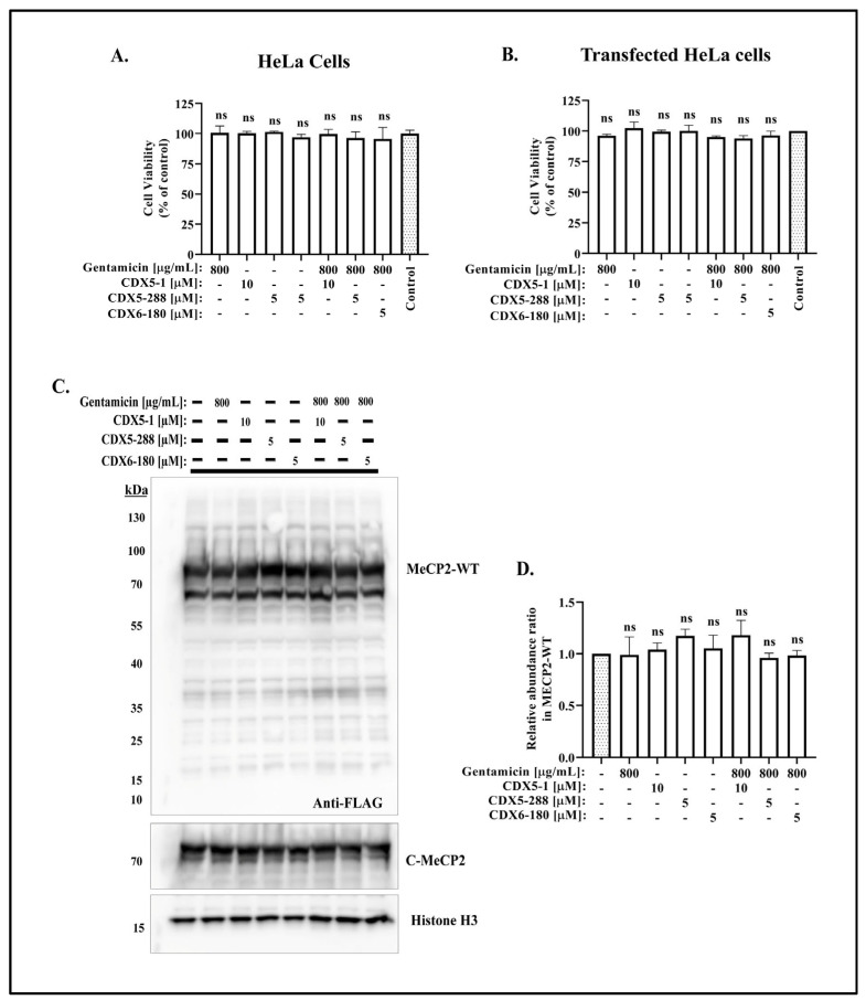
Rett syndrome (RTT), a severe X-linked neurodevelopmental disorder, is primarily caused by mutations in the methyl CpG binding protein 2 gene (MECP2). Over 35% RTT patients carry nonsense mutation in MECP2, making it a suitable candidate disease for nonsense suppression therapy. In our previous study, gentamicin was found to induce readthrough of MECP2 nonsense mutations with modest efficiency. Given the recent discovery of readthrough enhancers, CDX compounds, we herein evaluated the potentiation effect of CDX5-1, CDX5-288, and CDX6-180 on gentamicin-mediated readthrough efficiency in transfected HeLa cell lines bearing the four most common MECP2 nonsense mutations. We showed that all three CDX compounds potentiated gentamicin-mediated readthrough and increased full-length MeCP2 protein levels in cells expressing the R168X, R255X, R270X, and R294X nonsense mutations. Among all three CDX compounds, CDX5-288 was the most potent enhancer and enabled the use of reduced doses of gentamicin, thus mitigating the toxicity. Furthermore, we successfully demonstrated the upregulation of full-length Mecp2 protein expression in fibroblasts derived from Mecp2R255X/Y mice through combinatorial treatment. Taken together, findings demonstrate the feasibility of this combinatorial approach to nonsense suppression therapy for a subset of RTT patients.

Keywords: Rett syndrome; aminoglycosides; methyl-CpG-binding protein; nonsense suppression therapy; readthrough.


Conflict of interest statement

The authors declare no conflict of interest.

Figures

Figure 1
Figure 1
Enhancer CDX5-1 increases the efficiency of gentamicin-mediated readthrough. (A) Schematic representation of the FLAG-tagged MeCP2 highlighting the position of the mutants used in this study (NTD, N-terminal domain; MBD, methyl binding domain; ID, internal domain; TRD-NLS, transcription repression domain-nuclear localization signals; CTD, C-terminal domain). Western blot analysis of (B) R168X, (C) R255X, (D) R270X, and (E) R290X transfected HeLa cells treated with 800 µg/mL gentamicin and the indicated concentrations of CDX5-1 for 24 h, and probed with the indicated antibodies. The relative fold change of translational readthrough was calculated by comparing the expression of full-length MeCP2 in co-treatment to the expression of full-length MeCP2 in gentamicin treatment alone. Data are given as means ± SD, n ≥ 3 independent experiments. Statistical evaluation was performed using one-way ANOVA with Dunnett’s multiple comparison test.
Figure 2
Figure 2
Enhancer CDX5-288 increases the efficiency of gentamicin-mediated readthrough. Western blot analysis of (A) R168X, (B) R255X, (C) R270X, and (D) R290X transfected HeLa cells treated with 800 µg/mL gentamicin and the indicated concentrations of CDX5-288 for 24 h, and probed with the indicated antibodies. The relative fold change of translational readthrough was calculated by comparing the expression of full-length MeCP2 in co-treatment to the expression of full-length MeCP2 in gentamicin treatment alone. Data are given as means ± SD, n ≥ 3 independent experiments. Statistical evaluation was performed using one-way ANOVA with Dunnett’s multiple comparison test.
Figure 3
Figure 3
Enhancer CDX6-180 increases the efficiency of gentamicin-mediated readthrough. Western blot analysis of (A) R168X, (B) R255X, (C) R270X, and (D) R290X transfected HeLa cells treated with 800 µg/mL gentamicin and the indicated concentrations of CDX6-180 for 24 h, and probed with the indicated antibodies. The relative fold change in translational readthrough was calculated by comparing the expression of full-length MeCP2 in co-treatment to the expression of full-length MeCP2 in gentamicin treatment alone. Data are given as means ± SD, n ≥ 3 independent experiments. Statistical evaluation was performed using one-way ANOVA with Dunnett’s multiple comparison test.
Figure 4
Figure 4
Co-treatment of CDX compounds with gentamicin did not lead to cytotoxicity. Cell viability data for (A) HeLa cells and (B) FLAG-MeCP2-WT transfected HeLa cells treated with DMSO (control) or indicated concentration of gentamicin and CDX compounds for 24 h. Cell viability was calculated as % of control, and the effect of drug treatment was compared to control. (C) Western blot analysis of FLAG-MeCP2-WT transfected HeLa cells treated with indicated concentrations of gentamicin and CDX compounds for 24 h, and probed with the indicated antibodies. (D) Quantitative analysis of MeCP2 protein expression after 24 h of treatment. The relative abundance ratio was calculated by comparing the expression of MeCP2 protein in treated cells to the expression of MeCP2 protein in untreated cells, using FLAG densitometric readings. Data are given as means ± SD, n = 3 independent experiments. Statistical evaluation was performed using one-way ANOVA with Dunnett’s multiple comparison test. Note that not significant (ns) changes in all samples.
Figure 5
Figure 5
Nuclear localization of restored full-length MeCP2 protein. (A) Immunofluorescence studies of wild type-MeCP2, (B) R168X, (C) R255X, (D) R270X, and (E) R290X transfected HeLa cells treated with 800 µg/mL gentamicin and 5 µM of CDX5-288 or untreated. Staining of the Anti-FLAG (green signal) and Anti-C-MeCP2 (red signal) correspond with the DAPI nuclei staining (blue signal), thus indicating its nuclear localization (arrow). Scale bars: 20 µm.
Figure 6
Figure 6
CDX5-288 allows the use of reduced doses of gentamicin. Western blot analysis of R270X transfected HeLa cells that are treated with indicated concentration of gentamicin with or without (A) CDX5-1, (B) CDX5-288, (C) CDX6-180 for 24 h and probed with indicated antibodies. Right: quantitative analysis of full-length MeCP2 protein expression after 24 h of treatment. The relative readthrough translation (%) was calculated by comparing the expression of full-length MeCP2 in the treated samples to the expression of wild-type MeCP2, using the FLAG densitometric readings. The enhancement ratio represents the expression of full-length MeCP2 in cells co-treated with CDX compound and gentamicin relative to the expression of full-length MeCP2 in cells only treated with 800 µg/mL gentamicin alone. Data are given as means ± SD, n ≥ 3 independent experiments. Statistical evaluation was performed using one-way ANOVA with Dunnett’s multiple comparison test.
Figure 7
Figure 7
Combinatorial treatment enhances MeCP2 protein expression in Mecp2R255X/Y mouse-derived fibroblasts. (A) Western blot demonstrating no full-length MeCP2 protein from fibroblasts derived from Mecp2 R255X/Y mice, but full-length MeCP2 protein after 4 d treatment with 800 µg/mL gentamicin alone or in combination with CDX5-288. Each lane corresponds to an individual treatment experiment. (B) Quantitative analysis of MeCP2 protein expression in Mecp2R255X/Y mouse-derived fibroblasts. Data are given as means ± SD. Statistical evaluation was performed using unpaired Welch’s t-test.

References

    1. Rett A. On a unusual brain atrophy syndrome in hyperammonemia in childhood. Wien. Med. Wochenschr. 1966;116:723–726. - PubMed
    1. Hagberg B., Aicardi J., Dias K., Ramos O. A progressive syndrome of autism, dementia, ataxia, and loss of purposeful hand use in girls: Rett’s syndrome: Report of 35 cases. Ann. Neurol. 1983;14:471–479. doi: 10.1002/ana.410140412. - DOI - PubMed
    1. Huppke P., Held M., Laccone F., Hanefeld F. The spectrum of phenotypes in females with Rett Syndrome. Brain Dev. 2003;25:346–351. doi: 10.1016/S0387-7604(03)00018-4. - DOI - PubMed
    1. Neul J.L., Kaufmann W.E., Glaze D.G., Christodoulou J., Clarke A.J., Bahi-Buisson N., Leonard H., Bailey M.E.S., Schanen N.C., Zappella M., et al. Rett syndrome: Revised diagnostic criteria and nomenclature. Ann. Neurol. 2010;68:944–950. doi: 10.1002/ana.22124. - DOI - PMC - PubMed
    1. Amir R.E., Van den Veyver I.B., Wan M., Tran C.Q., Francke U., Zoghbi H.Y. Rett syndrome is caused by mutations in X-linked MECP2, encoding methyl-CpG-binding protein 2. Nat. Genet. 1999;23:185–188. doi: 10.1038/13810. - DOI - PubMed