Review
. 2023 Jul 20;24(14):11691.
doi: 10.3390/ijms241411691.

The Involvement of Polyunsaturated Fatty Acids in Apoptosis Mechanisms and Their Implications in Cancer

Affiliations
Review

The Involvement of Polyunsaturated Fatty Acids in Apoptosis Mechanisms and Their Implications in Cancer

Mayra Montecillo-Aguado et al. Int J Mol Sci. .

Abstract

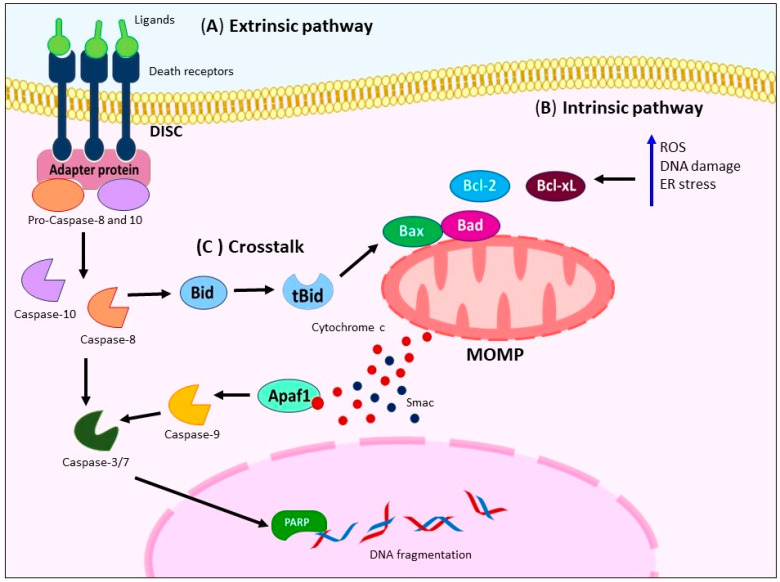
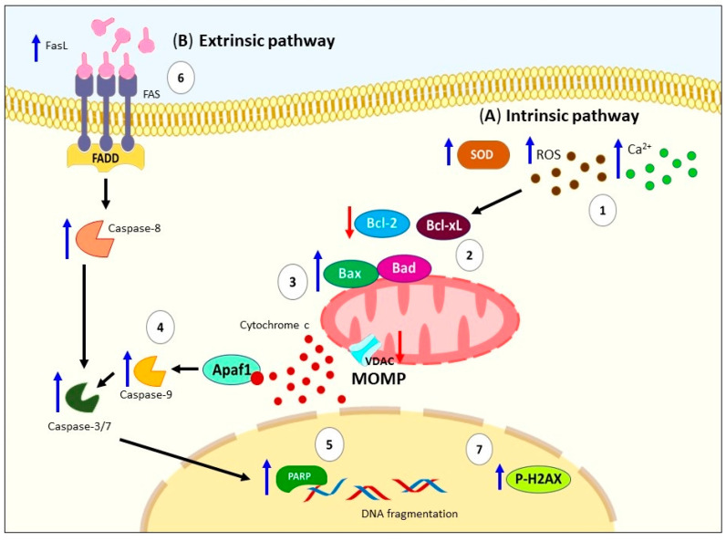
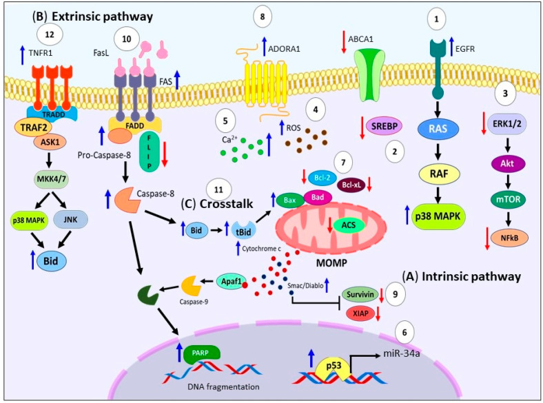
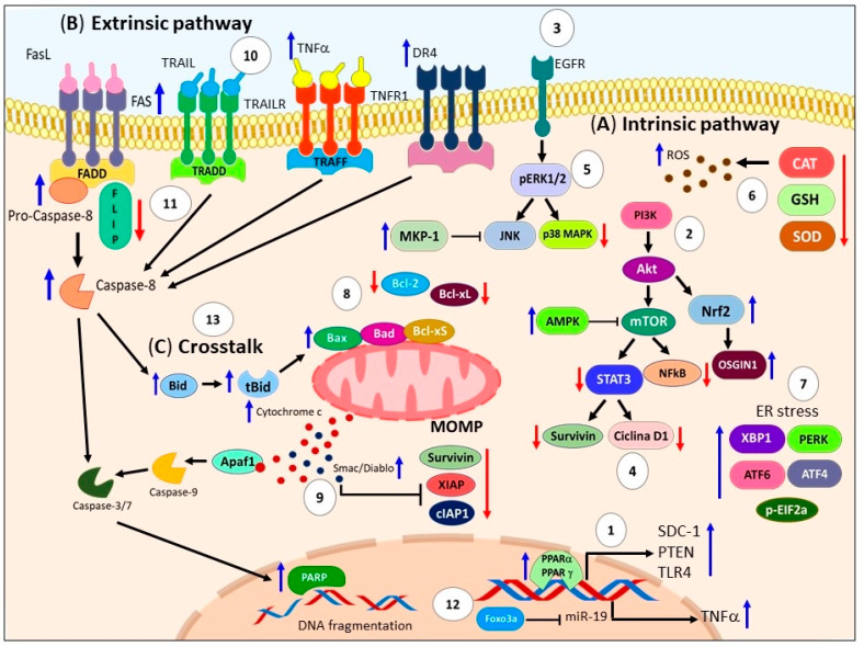
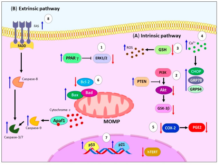
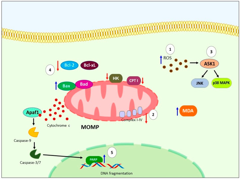
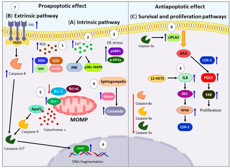
Cancer is a significant global public health issue and, despite advancements in detection and treatment, the prognosis remains poor. Cancer is a complex disease characterized by various hallmarks, including dysregulation in apoptotic cell death pathways. Apoptosis is a programmed cell death process that efficiently eliminates damaged cells. Several studies have indicated the involvement of polyunsaturated fatty acids (PUFAs) in apoptosis, including omega-3 PUFAs such as alpha-linolenic acid, docosahexaenoic acid, and eicosapentaenoic acid. However, the role of omega-6 PUFAs, such as linoleic acid, gamma-linolenic acid, and arachidonic acid, in apoptosis is controversial, with some studies supporting their activation of apoptosis and others suggesting inhibition. These PUFAs are essential fatty acids, and Western populations today have a high consumption rate of omega-6 to omega-3 PUFAs. This review focuses on presenting the diverse molecular mechanisms evidence in both in vitro and in vivo models, to help clarify the controversial involvement of omega-3 and omega-6 PUFAs in apoptosis mechanisms in cancer.

Keywords: apoptosis; cancer; polyunsaturated fatty acids (PUFAs).


Conflict of interest statement

The authors declare no conflict of interest.

Figures

Figure 1
Figure 1
Principal mechanism of apoptosis activation. (A) Activation of extrinsic pathway occurs through ligand binding to death receptors (FAS, TRAIL-R, or TNFR1), subsequently mediates bind of adapter proteins (FADD or TRADD) building DISC, activating caspase-8/-10 and -3. (B) Several stimuli trigger intrinsic pathway like cytokine deprivation, increase generation of ROS, DNA damage, and ER stress (blue arrow), causing MOMP that leads to the release of the mitochondrial proteins [cytochrome c (red dots), Smac (blue dots)], and subsequent activation of initiator caspase-9 and executioners caspases-3, -6, and -7 concluding in apoptosis. (C) There is crosstalk between the extrinsic and intrinsic pathways through the cleavage of caspase-8, which generates a tBid. tBid triggers MOMP, amplifying the apoptotic signal. FAS: Fas cell surface death receptor. TRAIL-R: TNF-related apoptosis-inducing ligand receptor. TNFR1: TNF receptor superfamily member 1A. FADD: FAS-associated death domain protein. TRADD: TNFR-associated death domain protein. DISC: Death-inducing signaling complex. ROS: Reactive oxygen species. DNA: Deoxyribonucleic acid. ER: Endoplasmic reticulum. MOMP: Mitochondrial outer membrane permeabilization. tBid: Truncated form of Bid.
Figure 2
Figure 2
Schematic representation of apoptosis activation by ALA. (A) Several signals can trigger the activation of intrinsic pathway by ALA, for example: (1) generation of ROS, induction of SOD activity, and increase in intracellular Ca2+; (2) these signals promote mitochondrial depolarization, downregulation of antiapoptotic proteins (Bcl-xL and Bcl-2); (3) upregulation of proapoptotic proteins (Bad and Bax), VDAC, Apaf, as well as cytochrome c release; (4) proteolytic activation of caspases -9 and -3; and (5) proteolytic cleavage of PARP. (B) Regarding the extrinsic pathway, there has been reported (6) high levels of FasL, activation of caspases-8, -3, -7, and DNA damage caused by (7) p-H2A.X. ALA: Alpha-linolenic acid. ROS: Reactive oxygen species. SOD: Superoxide dismutase. Bcl-xL: B-cell lymphoma-extra-large. Bcl-2: B-cell lymphoma 2. Bad: Bcl-2 associated agonist of cell death. Bax: Bcl-2 Associated X-protein. VDAC: Voltage-dependent anion channel. PARP: Poly(ADP-ribose) polymerase. FasL: Fas ligand. DNA: Deoxyribonucleic acid. P-H2A.X: Phospho-Histone H2A.X. MOMP: Mitochondrial outer membrane permeabilization.
Figure 3
Figure 3
Molecular pathways of EPA-induced apoptosis in cancer models. (A) EPA can trigger intrinsic pathway activation through arresting the progression of the cell cycle, (1) upregulation of Raf/MAPK pathway, sustained activation of EGFR/p38 MAPK axis, (2) accumulation of cholesterol by inhibition of SREBP2 and ABCA1, (3) downregulation of survival pathways (ERK1/2/Akt/mTOR/NFkB), (4) generation of ROS, (5) intracellular Ca2+ accumulation, and (6) Bcl-2 suppression through the p53/miR-34a axis, wt-p53 accumulation, activation of ACS. (7) These signals promote mitochondrial depolarization, and cytochrome c and Smac/Diablo release to the cytosol, activation of caspases -9, -3, -7, cleavage PARP, and DNA fragmentation. (8) Also, EPA causes upregulation of Bax, Bak and ADORA1. (9) In contrast, EPA promote downregulation of Bcl-xL, Survivin and XIAP. (B) Regarding the extrinsic pathway, (10) EPA increased FAS surface expression, downregulate FLIP levels, and promote activation of caspase-8. (C) (11) Numerous studies demonstrate crosstalk between both pathways through caspase-8 cleaved that generates tBid, which can be oligomerized with Bax and that triggers MOMP. Moreover, (12) treatment with C20E through TNFR1 activate ASK1-MKK4/7-JNK/p38MAPK pathway, promoting the tBid. EPA: Eicosapentanoic acid. Raf: Rapidly accelerated fibrosarcoma. MAPK: Mitogen-activated protein kinase. EGFR: Epidermal growth factor receptor. SREBP2: Cholesterol biosynthesis inducer. ABCA1: Cholesterol efflux channel protein. ERK1/2: Extracellular signal-regulated protein kinases 1 and 2. Akt: Protein kinase B. mTOR: Mammalian target of rapamycin. NFkB: Nuclear factor κB. ROS: Reactive oxygen species. Bcl-2: B-cell lymphoma 2. p53: Tumor protein P53. miR-34a: MicroRNA 34a. wt-p53: Wild type Tumor protein P53. ACS: Acyl-CoA synthetase. PARP: Poly(ADP-ribose) polymerase. DNA: Deoxyribonucleic acid. Bax: Bcl-2 Associated X-protein. Bak: Bcl-2 homologous antagonist/killer. ADORA1: Adenosine A1 Receptor. Bcl-xL: B-cell lymphoma-extra-large. XIAP: X-linked inhibitor of apoptosis protein. FAS: Fas cell surface death receptor. FLIP: FLICE inhibitory protein. tBid: Truncated form of Bid. MOMP: Mitochondrial outer membrane permeabilization. C20E: 17,18-epoxyeicosanoic acid. TNFR1: TNF receptor superfamily member 1A. ASK1: Apoptosis signal-regulating kinase 1. MKK4/7: MAP2K4 mitogen-activated protein kinase kinase 4. JNK: c-Jun N-terminal kinase.
Figure 4
Figure 4
Illustrates the apoptosis mechanisms triggered by DHA. (A) DHA induces modifications in the plasma membrane environment through various pathways. (1) It increases the expression of SDC-1 via PPARγ, (2) inhibits the Akt-mTOR axis by upregulating PPARγ and PTEN, downregulates the Akt/NFκB cell survival axis, (3) EGFR/STAT3/cyclin D1/survivin and (4) NF-κB/IκBα/cyclin D1/survivin pathways, (5) activates the ERK/JNK/p38 axis, stimulates AMPK, and downregulates the PI3K/Akt pathway. (6) EPA also leads to increased ROS production, activates the PI3K/Akt/Nrf2 signaling pathway, and induces the expression of OSGIN1. The downregulation of antioxidant enzymes such as CAT, GSH, and SOD contributes to the accumulation of intracellular ROS. Furthermore, (7) EPA activates ER stress through the expression of XBP1, PERK, ATF4, ATF6, and phosphorylated EIF2a. These events collectively induce intrinsic apoptosis by triggering the depolarization of the mitochondrial membrane. (8) This is achieved through the downregulation of Bcl-xL and Bcl-2, upregulation of Bax and Bcl-xS, and dimerization of Bax and Bak (9) Consequently, MOMP occurs, leading to the release of Smac/Diablo and cytochrome c from the mitochondria. Activated caspase-9 and -3/7 further execute apoptosis by cleaving PARP and forming DNA adducts. DHA downregulates XIAP, cIAP1, and survivin. (B) On the other hand, (10) DHA triggers the extrinsic pathway of apoptosis by increasing the expression of death receptors such as TRAIL, TNFR, DR4, and FAS. Subsequently, (11) DHA activates FADD and downregulates FLIP. Moreover, DHA sensitizes the apoptotic response to death ligands such as TNF-α, FAS antibody, and TRAIL. Additionally, (12) DHA induces nuclear accumulation of Foxo3a, which binds to the miR-21 promoter, leading to its transcriptional repression and increased TNFα mRNA levels. These events ultimately activate caspase 8 and executioner caspases, promoting apoptosis. (C) (13) Both EPA and DHA demonstrate crosstalk between the intrinsic and extrinsic apoptotic pathways through tBid. These findings highlight the multifaceted apoptotic mechanisms induced by DHA, involving both direct and indirect pathways, and emphasize its potential as a safe and effective option for cancer treatment. DHA: Docosahexaenoic acid. SDC-1: Syndecan-1. PPARγ: Peroxisome proliferator-activated receptor-gamma. Akt: Protein kinase B. mTOR: Mammalian target of rapamycin. PTEN: Phosphatase and tensin homolog. NFκB: Nuclear factor κB. EGFR: Epidermal growth factor receptor. STAT3: Signal transducer and activator of transcription 3. IκBα: Nuclear factor of kappa light polypeptide gene enhancer in B-cells inhibitor, alpha. ERK: Extracellular signal-regulated kinase. JNK: c-Jun N-terminal kinase. AMPK: AMP-activated protein kinase. PI3K: Phosphoinositide 3-kinase. ROS: Reactive oxygen species. Nrf2: Nuclear factor erythroid 2-related factor 2. OSGIN1: Oxidative stress-induced growth inhibitor 1. CAT: Catalase. GSH: Glutathione. SOD: Superoxide dismutase. ER: Endoplasmic reticulum. XBP1: X-box-binding protein 1. PERK: Protein kinase R-like endoplasmic reticulum kinase. ATF4: Activating Transcription Factor 4. ATF6: Activating Transcription Factor 6. EIF2a: Eukaryotic translation initiation factor 2A. Bcl-xL: B-cell lymphoma-extra-large. Bcl-2: B-cell lymphoma 2. Bax: Bcl-2 Associated X-protein. Bcl-xS: Bcl-2 homologous short isoform. Bak: Bcl-2 homologous antagonist/killer. MOMP: Mitochondrial outer membrane permeabilization. DNA: Deoxyribonucleic acid. XIAP: X-linked inhibitor of apoptosis protein. cIAP1: Cellular inhibitor of apoptosis protein 1. TRAIL: TNF-related apoptosis inducing ligand. TNFR: Epidermal growth factor receptor. DR4: Death receptor 4. FAS: Fas cell surface death receptor. FADD: FAS-associated death domain protein. FLIP: FLICE inhibitory protein. TNF-α: Tumor necrosis factor-alpha. Foxo3a: Forkhead transcription factor O subfamily member 3a. miR-21: MicroRNA 21. mRNA: Messenger ribonucleic acid. tBid: Truncated form of Bid.
Figure 5
Figure 5
Molecular pathways of apoptosis regulate by LA. (A) LA triggers mitochondrial pathway through inhibition of survival pathways, for example (1) downregulation of ERK1/2 by PPARγ, and (2) Akt/GSK-3β inhibition by PTEN activation. Moreover, (3) LA causes oxidative stress by massive ROS accumulation and GSH decrease. Likewise, (4) LA generates accumulation of intracellular Ca2+, and expression of the UPR-associated genes (CHOP, GRP78, and GRP94), (5) downregulation of the PGE2 production and telomerase activity through suppressing COX-2 and hTERT expression. (6) All these events cause loss of mitochondrial membrane potential, increase in the Bax and Bad expression, and downregulate expression of Bcl-2, producing MOMP and promote the release of cytochrome c from the mitochondria, causing activation of caspase-9 and caspase-3, and decreasing ATP level. (7) Additionally, other studies demonstrated the upregulation of p21 and p53 mRNA. (B) (8) Regarding extrinsic pathway LA increases FAS and caspase-8 levels. LA: Linoleic Acid. ERK1/2: Extracellular signal-regulated protein kinases 1 and 2. PPARγ: Peroxisome proliferator-activated receptor-gamma. Akt: Protein kinase B. GSK-3β: Glycogen Synthase Kinase 3 Beta. PTEN: Phosphatase and tensin homolog. ROS: Reactive oxygen species. GSH: Glutathione. UPR: Unfolded protein response. CHOP: C/EBP Homologous Protein. GRP78: Glucose-Regulated Protein 78. GRP94: Glucose-Regulated Protein 94. PGE2: Prostaglandin E2. COX-2: Cyclooxygenase 2. hTERT: Telomerase reverse transcriptase. Bax: Bcl-2 Associated X-protein. Bad: Bcl-2 associated agonist of cell death. Bcl-2: B-cell lymphoma 2. MOMP: Mitochondrial outer membrane permeabilization. ATP: Adenosine triphosphate. p21: Cyclin-dependent kinase inhibitor 1A. p53: Tumor protein P53. FAS: Fas cell surface death receptor.
Figure 6
Figure 6
Schematic representation of molecular apoptosis pathways regulates by GLA. (1) GLA causing oxidative stress by ROS accumulation and lipid peroxidation, additionally, (2) has been report downregulation of mitochondrial CPT I, hexoquinase, and mitochondrial respiratory chain complexes I+III and IV, high levels of MDA. (3) Another mechanism reported is ROS/ASK1/JNK/p38 MAPK pathway. (4) All these events trigger intrinsic apoptosis by downregulation of Bcl-Xl, Bcl-2, upregulation of Bad, causing loss of mitochondrial membrane potential, and subsequent release of cytochrome c, activation of caspase-9 and -3, (5) leading to cleavage of PARP and DNA fragmentation. GLA: Gamma-linolenic acid. ROS: Reactive oxygen species. CPT I: carnitine palmitoyltransferase I. MDA: malondialdehyde. ASK1: Apoptosis signal-regulating kinase 1. JNK: c-Jun N-terminal kinase. P38 MAPK: p38 MAP Kinase. Bcl-Xl: B-cell lymphoma-extra-large. Bcl-2: Bad: Bcl-2 associated agonist of cell death. PARP: Poly (ADP-ribose) polymerase. DNA: Deoxyribonucleic acid.
Figure 7
Figure 7
Effects of ARA on apoptotic death pathways. (A) (1) Treatment with ARA induces an increase in oxidative stress through the production of ROS and lipid peroxidation. This leads to elevated levels of MDA, 4-HNE, SOD, and GSH-PX activity. (2) Furthermore, the elevated ROS levels and intracellular calcium concentration activate the p38α MAPK and JNK1 pathways. (3) ARA also triggers ER stress, characterized by high levels of pXBP1 and phosphorylated eIF2α (p-eIF2α). (4) Another mechanism involves the accumulation of unmetabolized ARA, which activates the enzyme SMase, resulting in elevated ceramide levels—an important second messenger and potent activator of apoptosis. (5) These mechanisms collectively lead to the association of Bax protein with mitochondria, downregulation of Bcl-xL and Bcl-2, loss of mitochondrial membrane potential, release of cytochrome c from the mitochondria to the cytosol, and activation of caspase-9 and -3. (6) This cascade ultimately results in the breakdown of PPAR and DNA fragmentation. (B) (7) In the extrinsic pathway, ARA increases the expression of FAS, caspases-8, and -3. (C) On the other hand, ARA induces anti-apoptotic effects by activating survival and proliferation pathways, such as the Akt axis. (8) Additionally, 12-HETE triggers the activation of the ILK/NF-κB axis. (9) Another novel mechanism suggests that during apoptosis, active caspase-3 can activate ciPLA2, leading to the release of ARA and the production of PGE2. This abnormal activation of FAK promotes proliferation. Furthermore, downregulation of active caspases 3, 8, and 9 has been reported. ARA: Arachidonic acid. ROS: Reactive oxygen species. MDA: Malondialdehyde. 4-HNE: 4-hydroxy-2-nonenal. SOD: superoxide dismutase. GSH-PX: Glutathione peroxidase. p38α MAPK: p38α MAP Kinase. JNK1: c-Jun N-terminal kinase 1. ER: endoplasmic reticulum. pXBP1: processed form of XBP1. p-eIF2α: phosphorylated eukaryotic initiation factor-2α. SMase: sphingomyelinase. Bcl-xL: B-cell lymphoma-extra-large. Bcl-2: B-cell lymphoma 2. PPAR: Poly (ADP-ribose) polymerase. DNA: deoxyribonucleic acid. FAS: Fas cell surface death receptor. Akt: Protein kinase B. 12-HETE: 12-Hydroxyeicosatetraenoic acid. ILK: integrin-linked kinase. NF-κB: nuclear factor κB. ciPLA2: calcium-independent phospholipase A2. PGE2: Prostaglandin E2. FAK: Focal adhesion kinase.

References

    1. International Agency for Research on Cancer . All Cancers—WHO 2018. Volume 876. International Agency for Research on Cancer; Lyon, France: 2018. p. 2.
    1. Mohammad R.M., Muqbil I., Lowe L., Yedjou C., Hsu H.Y., Lin L.T., Siegelin M.D., Fimognari C., Kumar N.B., Dou Q.P., et al. Broad targeting of resistance to apoptosis in cancer. Semin. Cancer Biol. 2015;35:S78–S103. doi: 10.1016/j.semcancer.2015.03.001. - DOI - PMC - PubMed
    1. Fleisher T.A. Apoptosis. Ann. Allergy. Asthma Immunol. 1997;78:245–250. doi: 10.1016/S1081-1206(10)63176-6. - DOI - PubMed
    1. Vega O.M., Abkenari S., Tong Z., Tedman A., Huerta-Yepez S. Omega-3 Polyunsaturated Fatty Acids and Lung Cancer: Nutrition or Pharmacology? Nutr. Cancer. 2020;73:541–561. doi: 10.1080/01635581.2020.1761408. - DOI - PubMed
    1. Montecillo-Aguado M., Tirado-Rodriguez B., Tong Z., Vega O.M., Morales-Martínez M., Abkenari S., Pedraza-Chaverri J., Huerta-Yepez S. Importance of the role of ω-3 and ω-6 polyunsaturated fatty acids in the progression of brain cancer. Brain Sci. 2020;10:381. doi: 10.3390/brainsci10060381. - DOI - PMC - PubMed