. 2023 Jul 20;24(14):11705.
doi: 10.3390/ijms241411705.

Thymocyte Development of Humanized Mice Is Promoted by Interactions with Human-Derived Antigen Presenting Cells upon Immunization

Affiliations

Thymocyte Development of Humanized Mice Is Promoted by Interactions with Human-Derived Antigen Presenting Cells upon Immunization

Takataro Fukuhara et al. Int J Mol Sci. .

Abstract

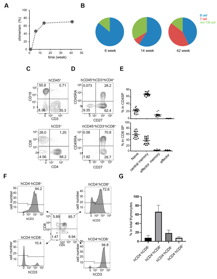
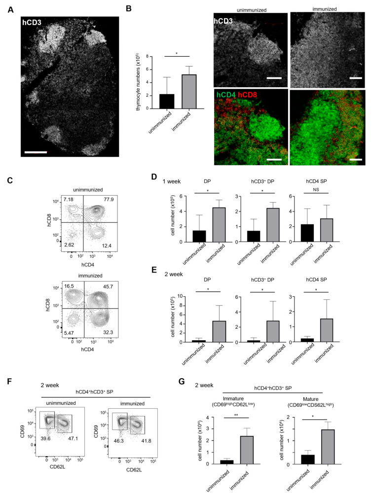
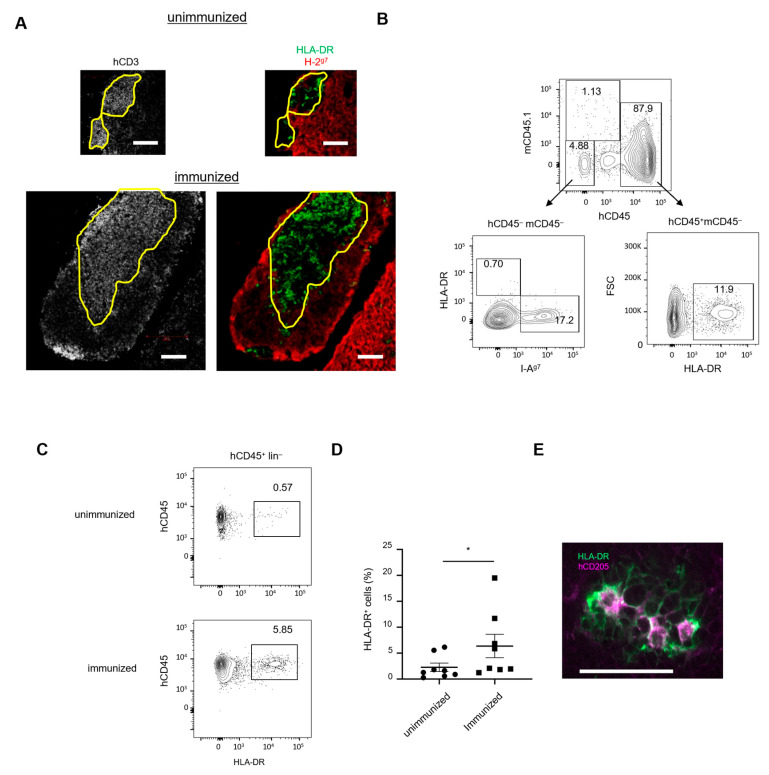
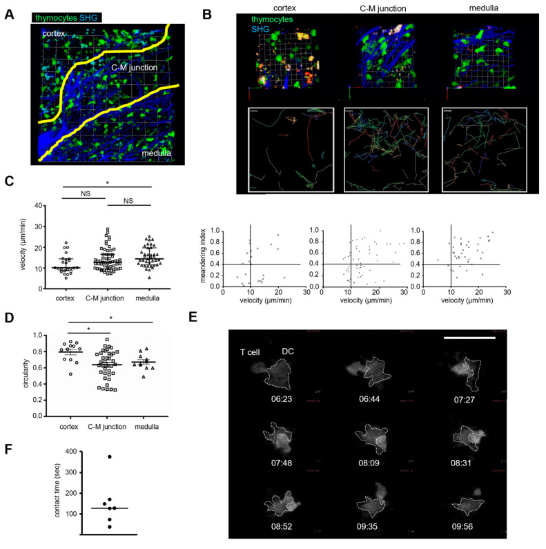
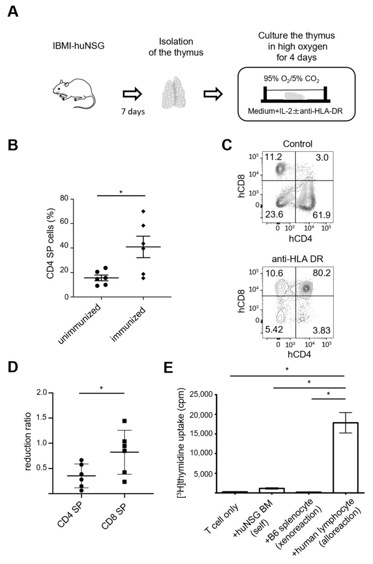
Immune responses in humanized mice are generally inefficient without co-transplantation of human thymus or HLA transgenes. Previously, we generated humanized mice via the intra-bone marrow injection of CD133+ cord blood cells into irradiated adult immunodeficient mice (IBMI-huNSG mice), which could mount functional immune responses against HTLV-1, although the underlying mechanisms were still unknown. Here, we investigated thymocyte development in IBMI-huNSG mice, focusing on the roles of human and mouse MHC restriction. IBMI-huNSG mice had normal developmental profiles but aberrant thymic structures. Surprisingly, the thymic medulla-like regions expanded after immunization due to enhanced thymocyte expansion in association with the increase in HLA-DR+ cells, including CD205+ dendritic cells (DCs). The organ culture of thymus from immunized IBMI-huNSG mice with a neutralizing antibody to HLA-DR showed the HLA-DR-dependent expansion of CD4 single positive thymocytes. Mature peripheral T-cells exhibited alloreactive proliferation when co-cultured with human peripheral blood mononuclear cells. Live imaging of the thymus from immunized IBMI-huNSG mice revealed dynamic adhesive contacts of human-derived thymocytes and DCs accompanied by Rap1 activation. These findings demonstrate that an increase in HLA-DR+ cells by immunization promotes HLA-restricted thymocyte expansion in humanized mice, offering a unique opportunity to generate humanized mice with ease.

Keywords: MHC; dendritic cells; humanized mice; immunization; thymus development.


Conflict of interest statement

The authors declare that they have no competing financial interest.

Figures

Figure 1
Figure 1
Generation of lymphocytes in IBMI-huNSG mice. (A) Typical time course of chimerism of human hematopoietic cells (human (h) CD45+ and mouse (m) CD45 cells) in the peripheral blood of IBMI-huNSG mice. (B) The proportion of B-cells (blue), T-cells (red), and non-T/B-cells (green) in hCD45+ cells in IBMI-huNSG mice. Weeks post-transplantation are indicated. (C) Representative flow cytometric profiles of human-derived peripheral T-cells (hCD3+) and B-cells (hCD19+) in IBMI-huNSG mice (14 weeks post-transplantation) (upper). Human CD4+ (hCD4+) and CD8+ T-cells (hCD8+) are also shown (below). (D) Flow cytometric profiles of hCD27 and hCD45 in CD4+ and CD8+ T-cells. (E) Fractions of naïve (hCD45RA+hCD27+, closed circle), memory (hCD45RAhCD27+, closed square), effector memory (hCD45RAhCD27, closed triangle), and terminal differentiated effector (hCD45RA+hCD27, closed inverted triangle) cells in CD4+ (upper) and CD8+ T-cells (lower). The bars represent average percentages (n = 14). (F) Thymocyte development in IBMI-huNSG mice. Expression of CD3 in DN (hCD4hCD8), DP (hCD4+hCD8+), CD4 SP (hCD4+hCD8), and CD8 SP (hCD4hCD8+) thymocytes of IBMI-huNSG mice (14 weeks). (G) The frequency of thymocyte subsets of IBMI-huNSG mice (n = 10).
Figure 2
Figure 2
Enhanced thymocyte development in enlarged medulla-like regions of the thymus from humanized mice after immunization. (A) Representative images of the thymus stained with anti-hCD3 antibody show focal aggregation of developed thymocyte near the capsule in IBMI-huNSG mice (28 weeks). Bar, 200 μm. (B) Total thymocyte numbers before and 1 week after immunization (left). Averages with SEM are shown (unimmunized, n = 5; immunized, n = 5). Representative images of the thymus from unimmunized and immunized IBMI-huNSG mice stained with antibodies against hCD3, hCD4 (green), and hCD8 (red). Bars represent 100 μm. (C) Representative flowcytometric profiles of hCD4 and hCD8 in the thymus from unimmunized and immunized IBMI-huNSG mice (day 7). (D,E) Changes in cell numbers of DP, CD3+ DP, and CD4 SP thymocytes with or without immunization. Cell numbers 1 week (unimmunized, n = 5; immunized, n = 5) (D) and 2 weeks after immunization (unimmunized, n = 5; immunized, n = 6) (E) are shown. Averages with SEM are shown. (F) hCD69 and hCD62L expression profiles of hCD4 SP cells measured by flow cytometry. The gate strategy and percentage of CD69highCD62Llow and CD69lowCD62Lhigh are shown and represent immature and mature subsets, respectively. (G) Changes in cell numbers of CD69highCD62Llow and CD69lowCD62Lhigh CD4 SP thymocytes with or without immunization. Cell numbers at 2 weeks (unimmunized, n = 5; immunized, n = 6). The statistical significance of the above data (D,E,G) was calculated by Student’s t-test. * p < 0.05, ** p < 0.01, NS: not significant.
Figure 3
Figure 3
Recruitment of HLA-DR+ DCs in the thymus after immunization. (A) Distribution of human HLA-DR and mouse MHC class II in the thymus. Frozen sectioned thymic tissues from immunized IBMI-huNSG mice were stained with antibodies against hCD3 (left images) or HLA-DR (green) and I-Ag7 (red) (right images). Bars represent 100 μm. (B) Flow cytometric profiles of hematopoietic or non-hematopoietic cells in the thymus from IBMI-huNSG mice (upper). Non-hematopoietic cells gated for hCD45 mCD45.1 contained the I-Ag7+ population (17.2%) but few HLA-DR+ cells (0.7%) (lower left), whereas human-derived hematopoietic cells (hCD45+ mCD45) contained the HLA-DR+ population (11.9%) (lower right). (C) HLA-DR expression in the human hematopoietic cells negative for the lineage markers hCD3 (T-cells), hCD20 (B-cells), hCD14 (monocytes/macrophages), and hCD56 (NK cells), with/without immunization. (D) Percentages of the HLA-DR+ in hCD45+lin cells with/without immunization (unimmunized, n = 8; immunized, n = 8). Bars represent averages with SEM of the populations. Statistical significance was determined by Mann–Whitney U test (* p < 0.05). (E) Immunostaining for HLA-DR (green) and CD205 (red) in thymus from immunized IBMI-huNSG mice. Bar represents 50 μm.
Figure 4
Figure 4
Two-photon imaging of human thymocyte–DC interactions in the thymus. (A) Two-photon live imaging of the thymus from IBMI-huNSG mice transplanted with CD133+ HSCs expressing Venus. Most Venus-expressing cells are morphologically identified as thymocytes and DCs. Cortex, medulla, and cortico-medullary junction rich in fibrous collagen (blue) are indicated. One square unit represents 25 μm × 25 μm. The data are representative of four experiments. (B) Comparison of cell motilities of thymocytes in the cortex, cortico-medullary junction, and medulla. Representative 3D reconstituted images of each region of the thymus (upper, 1 × 1 unit = 7.5 μm × 7.5 μm) and trajectories of thymocytes (middle, a scale bar, 5.1 μm) are shown. The plot of thymocyte velocities (μm/min) and meandering index of trajectories (straightness) in each region is also shown (bottom). (C) Thymocyte velocities (μm/min) in the cortex, cortico-medullary junction, and medulla. (D) Comparison of thymocyte circularity in the cortex, cortico-medullary junction, and medulla. Bars represent averages and SEM. (E) A representative image sequence of thymocyte–DC interactions is shown with time (min: s). Thymocyte and DCs (marked with dashed lines) express a Rap affinity probe. Bar represents 30 μm. The data are representative of two experiments. (F) Contact duration of thymocyte–DC interactions (n = 7). Statistical significance above data (C,D) by unpaired Student t-test are indicated: * p < 0.05, NS: not significant).
Figure 5
Figure 5
HLA-DR-dependent thymocyte maturation and allogeneic response of T-cells. (A) Schema of organ culture of the thymus. Thymic lobes were isolated 7 days after immunization of IBMI-huNSG mice and cultured in the absence or presence of anti-HLA-DR antibodies. Organ culture was supplemented with IL-2 and high oxygen. (B) Percentages of CD4 SP thymocytes in cultured thymus from humanized mice with/without immunization (unimmunized, n = 6; immunized, n = 6). Bars are averages with SEM. (C,D) Anti-HLA-DR antibody inhibited the generation of CD4 SP thymocytes. Representative flow cytometric profile (C) and reduction ratios of CD4 and CD8 SP thymocytes normalized against control antibodies (unimmunized, n = 6; immunized, n = 6). Bars represent averages with SEM of the populations. (E) Mixed lymphocyte reaction of peripheral T-cells from immunized IBMI-huNSG mice using splenic T-cells from syngeneic bone marrow cells from huNSG mice (huNSG BM), xenogeneic splenocytes from C57BL/6 mice, and allogeneic human-derived peripheral lymphocytes. Proliferation, as reflected by the uptake of [3H]-thymidine measured in triplicate (day 3), is shown. Averages with SEM are shown, with statistical significance (unpaired Student t-test; * p < 0.05). The data are representative of two experiments. Statistical significance above data (B,D,E) was determined by paired t-test: * p < 0.05.

Similar articles

References

    1. Ito R., Takahashi T., Ito M. Humanized mouse models: Application to human diseases. J. Cell Physiol. 2018;233:3723–3728. doi: 10.1002/jcp.26045. - DOI - PubMed
    1. Skelton J.K., Ortega-Prieto A.M., Dorner M. A Hitchhiker’s guide to humanized mice: New pathways to studying viral infections. Immunology. 2018;154:50–61. doi: 10.1111/imm.12906. - DOI - PMC - PubMed
    1. Walsh N.C., Kenney L.L., Jangalwe S., Aryee K.E., Greiner D.L., Brehm M.A., Shultz L.D. Humanized Mouse Models of Clinical Disease. Annu. Rev. Pathol. 2017;12:187–215. doi: 10.1146/annurev-pathol-052016-100332. - DOI - PMC - PubMed
    1. Rongvaux A., Takizawa H., Strowig T., Willinger T., Eynon E.E., Flavell R.A., Manz M.G. Human hemato-lymphoid system mice: Current use and future potential for medicine. Annu Rev. Immunol. 2013;31:635–674. doi: 10.1146/annurev-immunol-032712-095921. - DOI - PMC - PubMed
    1. Shultz L.D., Brehm M.A., Garcia-Martinez J.V., Greiner D.L. Humanized mice for immune system investigation: Progress, promise and challenges. Nat. Rev. Immunol. 2012;12:786–798. doi: 10.1038/nri3311. - DOI - PMC - PubMed

Substances