. 2023 Jul 21;24(14):11770.
doi: 10.3390/ijms241411770.

Genome-Wide Identification of Aqp Family Related to Spermatogenesis in Turbot (Scophthalmus maximus)

Affiliations

Genome-Wide Identification of Aqp Family Related to Spermatogenesis in Turbot (Scophthalmus maximus)

Xueying Wang et al. Int J Mol Sci. .

Abstract

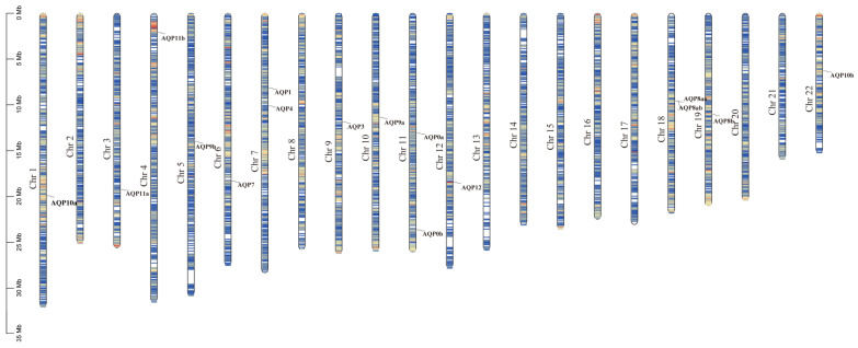
The development and maturation of sperm entails intricate metabolic processes involving water molecules, amino acids, hormones, and various substances. Among these processes, the role of aquaporins (aqps) in the testis is crucial. Turbot (Scophthalmus maximus) is a significant marine flatfish species in China; however, natural egg laying in females is not feasible under cultured conditions. Consequently, artificial insemination becomes necessary, requiring the retrieval of sperm and eggs through artificial methods. In this study, we combined genomic, transcriptomics, RT-qPCR, computer-assisted sperm analysis (CASA), and immunohistochemistry to investigate the involvement of the aqp family in spermatogenesis in turbot. Through genomic data analysis, we identified 16 aqps genes dispersed across 13 chromosomes, each exhibiting the characteristic major intrinsic protein (MIP) domain associated with AQPs. The results from RNA-seq and RT-qPCR analysis revealed prominent expression of aqp4, 10, and 12 during the proliferative stage, whereas aqp1 showed primary expression during the mature stage. aqp11 displayed high expression levels during both MSII and MSV stages, potentially contributing significantly to the proliferation and maturation of male germ cells. Conversely, aqp8 showed elevated expression levels during the MSIII, MSIII-IV, and MSIV stages, suggesting its direct involvement in spermiogenesis. Immunohistochemical analysis unveiled the predominant localization of AQP1 protein in male germ cells rather than Sertoli cells, specifically concentrated in the head of sperm within cysts. Furthermore, a noteworthy decline in sperm motility was observed when sperm were subjected to treatment with either the AQP1-specific inhibitor (HgCl2) or the AQP1 antibody. However, no direct correlation was found between the expression of Smaqp1 and sperm quality. Overall, these findings provide new insights into the involvement of aqps in teleost spermatogenesis. Moreover, they hold potential for improving techniques related to sperm activation and cryopreservation, offering valuable knowledge for future advancements in this field.

Keywords: aqps; sperm; spermatogenesis; turbot.


Conflict of interest statement

The authors declare that there is no conflict of interest.

Figures

Figure 1
Figure 1
Phylogenetic analysis of AQP amino acid sequences from ten species. Each branch is color-coded to group the same AQP from different species together. The species included in the analysis were Dr (Danio rerio), Sm (Scophthalmus maximus), Po (Paralichthys olivaceus), Ol (Oryzias latipes), Su (Sebastes umbrosus), Xm (Xiphophorus maculatus), Ss (Sebastes schlegelii), Hs (Homo sapiens), Mm (Mus musculus), and Gg (Gallus gallus). The diameter of the red circles represents the size of the bootstrap on the left side. The red circles of different diameters were on the phylogenetic tree branch on the right side.
Figure 2
Figure 2
Motif and conserved domain analysis of AQP protein. The diverse domains identified were represented by different colors in the rectangles. The first column was visualization of the evolutionary tree of family members. On the left side, the colored square blocks are motifs that are conserved among AQPs (the second column). On the right side, the colored square blocks are domains that are conserved among AQPs (the third column). The fourth column is the legend: the top is motif, and the bottom is domain.
Figure 3
Figure 3
Chromosome localization of aqp genes in S. maximus (Chr: chromosome).
Figure 4
Figure 4
The expression levels of aqps genes were analyzed using RNA-seq at various stages of testis development (p < 0.05). Panels (AF) depict the expression patterns of specific aqp genes. (A): aqp1; (B): aqp4; (C): aqp8; (D): aqp10; (E): aqp11; (F): aqp12. The developmental stages analyzed include MSII and MSIII, representing the initiation stage of the annual breeding cycle; MSIII–IV, indicating the transitional phase of the testes; MSIV, representing the spermiogenesis stage with an increased proportion of spermatid; MSV, corresponding to the spawning phase; and MSVI, representing the testis recession phase. According to statistical analysis, the letter “a” represents the relative expression levels whose mean values are relatively larger. From “a” to “c”, the mean values decrease in turn. There is no significant difference between stages with the same letter. There is a significant difference between stages with different letters, (p < 0.05).
Figure 5
Figure 5
The expression levels of aqps genes were analyzed at different testis development stages (p < 0.05). The results are presented as follows: (A): aqp1; (B): aqp4; (C): aqp8; (D): aqp10; (E): aqp11; (F): aqp12. The developmental stages analyzed include MSII and MSIII, representing the initiation stage of the annual breeding cycle; MSIII–IV, indicating the transitional phase of the testes; MSIV, representing the spermiogenesis stage with an increased proportion of spermatid; MSV, corresponding to the spawning phase; and MSVI, representing the testis recession phase. The ubq and rsp genes were used as reference genes. Relative gene expression data were analyzed using the 2−ΔΔCt method. All reactions were carried out in triplicate. According to statistical analysis, the letter “a” represents the relative expression levels whose mean values are relatively larger. From “a” to “d”, the mean values decrease in turn. There is no significant difference between stages with the same letter. There is a significant difference between stages with different letters, (p < 0.05).
Figure 6
Figure 6
The localization of AQP1 protein in testis during sperm maturation. (A) the histological structure of the testis during sperm maturation, visualized through HE staining; (B) the negative control; (C) the localization of the AQP1 protein in the testis, 10×; and (D) the localization of the AQP1 protein in the testis, 20×. The scale bar represents 100 μm. The arrowhead in (A) indicates the sperm cyst; arrows in (C) indicate somatic cells surrounding the germ cells, and arrows in (D) indicate sperm exhibiting positive signal.
Figure 7
Figure 7
The correlation between aqp1 expression and sperm motility. (A) Effects of aqp1 inhibitor and antibody on sperm motility (p < 0.05). (B) Expression of aqp1 across different sperm quality groups, categorized as low, middle and high sperm motility (p < 0.05). Motility parameters in (A,B) were motility rate. The motility range as low (<20%) to middle (40–60%) to high (80–90%) ranges in (B). According to statistical analysis, the letter “a” represents the relative expression levels whose mean values are relatively larger. From “a” to “c”, the mean values decrease in turn. There is no significant difference between stages with the same letter. There is a significant difference between stages with different letters, (p < 0.05).

References

    1. Alavi S.M.H., Cosson J. Sperm motility in fishes (II) Effects of ions and osmolality: A review. Cell Biol. Int. 2006;30:1–14. doi: 10.1016/j.cellbi.2005.06.004. - DOI - PubMed
    1. Herrera F., Bondarenko O., Boryshpolets S. Osmoregulation in fish sperm. Fish Physiol. Biochem. 2021;47:785–795. doi: 10.1007/s10695-021-00958-1. - DOI - PubMed
    1. Cerda J., Finn R.N. Piscine aquaporins: An overview of recent advances. J. Exp. Zool. 2010;313A:623–650. doi: 10.1002/jez.634. - DOI - PubMed
    1. King L.S., Kozono D., Agre P. From structure to disease: The evolving tale of aquaporin biology. Nat. Rev. Mol. Cell Biol. 2004;5:687–698. doi: 10.1038/nrm1469. - DOI - PubMed
    1. Zhang D., Tan Y.J., Qu F., Sheng J.Z., Huang H.F. Functions of water channels in male and female reproductive systems. Mol. Asp. Med. 2012;33:676–690. doi: 10.1016/j.mam.2012.02.002. - DOI - PubMed

LinkOut - more resources