. 2023 Jul 21;24(14):11773.
doi: 10.3390/ijms241411773.

Sugar Starvation Disrupts Lipid Breakdown by Inducing Autophagy in Embryonic Axes of Lupin (Lupinus spp.) Germinating Seeds

Affiliations

Sugar Starvation Disrupts Lipid Breakdown by Inducing Autophagy in Embryonic Axes of Lupin (Lupinus spp.) Germinating Seeds

Sławomir Borek et al. Int J Mol Sci. .

Abstract

Under nutrient deficiency or starvation conditions, the mobilization of storage compounds during seed germination is enhanced to primarily supply respiratory substrates and hence increase the potential of cell survival. Nevertheless, we found that, under sugar starvation conditions in isolated embryonic axes of white lupin (Lupinus albus L.) and Andean lupin (Lupinus mutabilis Sweet) cultured in vitro for 96 h, the disruption of lipid breakdown occurs, as was reflected in the higher lipid content in the sugar-starved (-S) than in the sucrose-fed (+S) axes. We postulate that pexophagy (autophagic degradation of the peroxisome-a key organelle in lipid catabolism) is one of the reasons for the disruption in lipid breakdown under starvation conditions. Evidence of pexophagy can be: (i) the higher transcript level of genes encoding proteins of pexophagy machinery, and (ii) the lower content of the peroxisome marker Pex14p and its increase caused by an autophagy inhibitor (concanamycin A) in -S axes in comparison to the +S axes. Additionally, based on ultrastructure observation, we documented that, under sugar starvation conditions lipophagy (autophagic degradation of whole lipid droplets) may also occur but this type of selective autophagy seems to be restricted under starvation conditions. Our results also show that autophagy occurs at the very early stages of plant growth and development, including the cells of embryonic seed organs, and allows cell survival under starvation conditions.

Keywords: asparagine; embryo; iTRAQ; lipid droplet; lipophagy; peroxisome; pexophagy; proteomics; transcriptomics; ultrastructure.


Conflict of interest statement

The authors declare no conflict of interest.

Figures
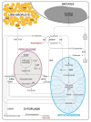
Figure 1
Figure 1
Simplified scheme of interconnections between storage lipid and protein breakdown in germinating lupin seeds. During lipid breakdown, fatty acids released by lipases from triacylglycerols (TAG) deposited in lipid droplets are oxidized in the peroxisome through β-oxidation. The next step of lipid breakdown involves the glyoxylate cycle, which operates partially in the peroxisome (also known as the glyoxysome) and the cytoplasm. The final product of lipid mobilization is sucrose, which is one of the most important carbon transport forms, and it can also be used as a respiratory substrate. However, in germinating lupin seeds, the main respiratory substrates are amino acids. With the intense amino acid interconversions in tissues of lupin germinating seeds, toxic ammonia is generated, and to maintain a low level of this byproduct, asparagine is synthesized in high amounts to utilize ammonia. Some metabolites of the glyoxylate cycle are subtracted from the typical pathway of lipid breakdown and can be used as respiratory substrates as well or be involved in amino acid metabolism. It was evidenced that in germinating lupine seeds, a pool of lipid-derived carbon skeletons is directed for amino acid synthesis, and glutamate and asparagine are among the main targets of carbon flow from lipid to amino acids. Based on data from the literature [2,4,9,10,11,12]. ACOX Acyl-CoA oxidase, AS asparagine synthase (glutamine-hydrolyzing), ATs aminotransferases, cACO aconitase (cytosolic fraction), cIDH isocitrate dehydrogenase (NADP-dependent, cytosolic fraction), cMDH malate dehydrogenase (cytosolic fraction), GDH glutamate dehydrogenase, GOGAT glutamate synthase, GS glutamine synthetase (glutamate-ammonia ligase), ICL isocitrate lyase, CAT catalase, LIP lipase, PEPCK phosphoenolpyruvate carboxykinase, TAG triacylglycerol.
Figure 2
Figure 2
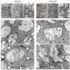
Ultrastructure of root meristematic zone cells of white and Andean lupin isolated embryo axes cultured in vitro for 96 h on a medium with (+S) and without (-S) 60 mM sucrose. Culture media were also enriched with 35 mM asparagine (+Asn). AB—autophagic body, CW—cell wall, LD—lipid droplet, N—nucleus, Nu—nucleolus, S—starch, SP—storage protein, V—vacuole. Scale bars (red; above the micrographs) = 2.0 μm.
Figure 3
Figure 3
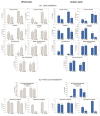
Total lipid content (a) and phospholipids (b) in white and Andean lupin isolated embryo axes cultured in vitro for 96 h on a medium with (+S) and without (-S) 60 mM sucrose. Culture media were also enriched with 35 mM asparagine (+Asn). Different letters above the error bars (±SD) indicate statistically significant differences at p ≤ 0.05 (ANOVA, Tukey’s HSD multiple-range test).
Figure 4
Figure 4
The activity of selected enzymes involved in lipid breakdown (a) and amino acid metabolism (b) in white and Andean lupin isolated embryo axes cultured in vitro for 96 h on a medium with (+S) and without (-S) 60 mM sucrose. Culture media were also enriched with 35 mM asparagine (+Asn). Different letters above the error bars (±SD) indicate statistically significant differences at p ≤ 0.05 (ANOVA, Tukey’s HSD multiple-range test).
Figure 5
Figure 5
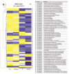
Heatmaps showing the relations in the level of transcripts of genes encoding proteins involved in central amino acid metabolism in white lupin (a) and Andean lupin (b) isolated embryonic axes cultured in vitro for 96 h on a medium with (+S) and without (-S) 60 mM sucrose. Culture media were enriched with 35 mM asparagine (+Asn). The data represent averages obtained from three independent experiments. The list of transcripts was made based on data from the literature [4,11,31] and the KEGG database dedicated to the narrow-leaved blue lupin (Lupinus angustifolius) genome (https://www.genome.jp/kegg-bin/show_organism?menu_type=pathway_maps&org=lang; accessed on 16 June 2022). The transcriptomics data selected for the preparation of this figure are presented in Table S2.
Figure 6
Figure 6
Heatmaps showing relations in transcript levels of genes encoding proteins involved in lipid degradation in white lupin (a) and Andean lupin (b) isolated embryonic axes cultured in vitro for 96 h on a medium with (+S) and without (-S) 60 mM sucrose. Culture media were also enriched with 35 mM asparagine (+Asn). The data represent averages obtained from three independent experiments. The list of transcripts was made based on data from the literature [2,9,10,11] and the KEGG database dedicated to the narrow-leaved blue lupin (Lupinus angustifolius) genome (https://www.genome.jp/kegg-bin/show_organism?menu_type=pathway_maps&org=lang; accessed on 14 June 2022). The transcriptomics data selected for the preparation of this figure are presented in Table S3.
Figure 6
Figure 6
Heatmaps showing relations in transcript levels of genes encoding proteins involved in lipid degradation in white lupin (a) and Andean lupin (b) isolated embryonic axes cultured in vitro for 96 h on a medium with (+S) and without (-S) 60 mM sucrose. Culture media were also enriched with 35 mM asparagine (+Asn). The data represent averages obtained from three independent experiments. The list of transcripts was made based on data from the literature [2,9,10,11] and the KEGG database dedicated to the narrow-leaved blue lupin (Lupinus angustifolius) genome (https://www.genome.jp/kegg-bin/show_organism?menu_type=pathway_maps&org=lang; accessed on 14 June 2022). The transcriptomics data selected for the preparation of this figure are presented in Table S3.
Figure 7
Figure 7
Heatmaps showing relations in levels of transcripts of genes encoding proteins involved in pexophagy (autophagic degradation of peroxisomes) in white lupin (a) and Andean lupin (b) isolated embryonic axes cultured in vitro for 96 h on a medium with (+S) and without (-S) 60 mM sucrose. Culture media were also enriched with 35 mM asparagine (+Asn). The data represent averages obtained from three independent experiments. The list of transcripts was made based on literature data [29,32,33,34]. Transcriptomics data selected for the preparation of this figure are presented in Table S4.
Figure 8
Figure 8
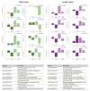
Relative changes in levels of selected gene transcripts determined by qRT-PCR technique in white and Andean lupin isolated embryonic axes cultured in vitro for 96 h on a medium with (+S) and without (-S) 60 mM sucrose. Culture media were also enriched with 35 mM asparagine (+Asn). Primer sequences used for qRT-PCR reactions are presented in Table S5. The comparative CT method for relative quantification was used with actin as an endogenous control. The amount of target, normalized to an endogenous reference and relative to the calibrator (+S; equal 1), is given by 2−ΔΔCT [35]. The data represent averages obtained from three independent experiments. Different letters above the error bars (±SD) indicate statistically significant differences at p ≤ 0.05 (ANOVA, Tukey’s HSD multiple-range test).
Figure 9
Figure 9
Western blot (a) and densitometric analysis (b) of the peroxisomal membrane protein Pex14 (Pex14p) content in white and Andean lupin isolated embryonic axes cultured in vitro for 96 h on a medium with (+S) and without (-S) 60 mM sucrose. Culture media were also enriched with 35 mM asparagine (+Asn). After 72 h of in vitro culture, for 24 h before the analysis, axes were transferred to media containing 10 μM concanamycin A (Con A), an inhibitor of autophagy (Con A inhibits the tonoplast PPi-dependent proton pump, causing neutralization of vacuolar pH, and thereby inhibits the activity of vacuolar hydrolytic enzymes, and in consequence, degradation of autophagic bodies inside the vacuole). The western blots presented in part (a) are representative of three independent experiments. Consistently, 40 μg of total protein was loaded per cell during electrophoresis. The data presented in part (b) are averages obtained from three independent experiments. The +S axes were set as 100% and were treated as a reference for axes from the rest of the trophic variants of the in vitro culture. Different letters above the error bars (±SD) indicate statistically significant differences at p ≤ 0.05 (ANOVA, Tukey’s HSD multiple-range test).

Similar articles

Cited by

References

    1. Ratajczak W., Borek S., Podgorski A., Ratajczak L. Variability of globulin composition in cultivars and individually tested seeds of yellow lupin (Lupinus luteus L.) Acta Physiol. Plant. 1999;21:413–417. doi: 10.1007/s11738-999-0014-z. - DOI
    1. Borek S., Ratajczak W., Ratajczak L. Regulation of storage lipid metabolism in developing and germinating lupin (Lupinus spp.) seeds. Acta Physiol. Plant. 2015;37:119. doi: 10.1007/s11738-015-1871-2. - DOI
    1. Borek S., Galor A., Paluch E. Asparagine Enhances Starch Accumulation in Developing and Germinating Lupin Seeds. J. Plant Growth Regul. 2013;32:471–482. doi: 10.1007/s00344-012-9313-5. - DOI
    1. Lehmann T., Ratajczak L. The pivotal role of glutamate dehydrogenase (GDH) in the mobilization of N and C from storage material to asparagine in germinating seeds of yellow lupine. J. Plant Physiol. 2008;165:149–158. doi: 10.1016/j.jplph.2006.12.010. - DOI - PubMed
    1. Gaufichon L., Reisdorf-Cren M., Rothstein S.J., Chardon F., Suzuki A. Biological functions of asparagine synthetase in plants. Plant Sci. 2010;179:141–153. doi: 10.1016/j.plantsci.2010.04.010. - DOI

LinkOut - more resources