. 2023 Jul 24;24(14):11842.
doi: 10.3390/ijms241411842.

Tea Polyphenols Protects Tracheal Epithelial Tight Junctions in Lung during Actinobacillus pleuropneumoniae Infection via Suppressing TLR-4/MAPK/PKC-MLCK Signaling

Affiliations

Tea Polyphenols Protects Tracheal Epithelial Tight Junctions in Lung during Actinobacillus pleuropneumoniae Infection via Suppressing TLR-4/MAPK/PKC-MLCK Signaling

Xiaoyue Li et al. Int J Mol Sci. .

Abstract

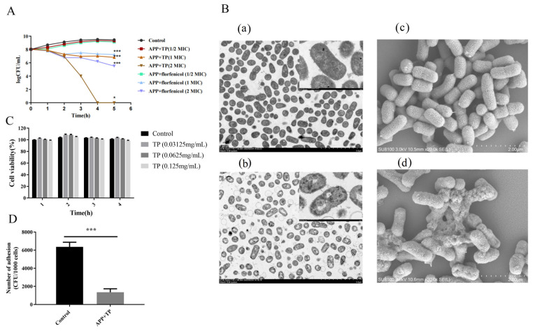
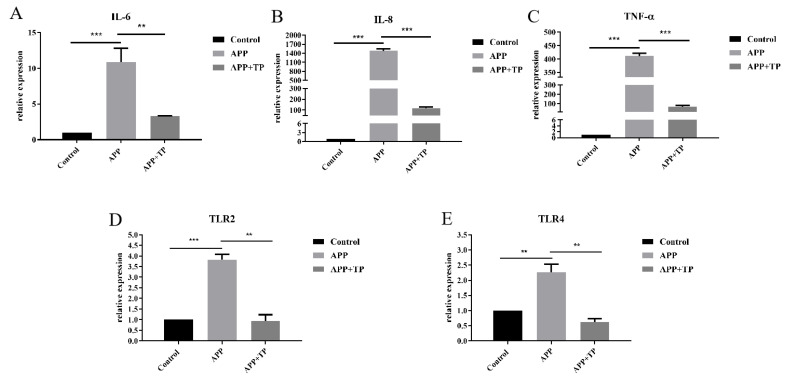
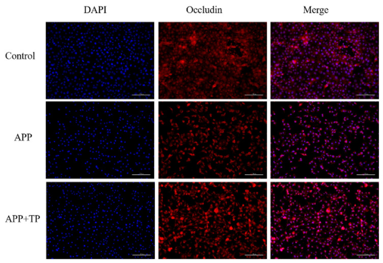
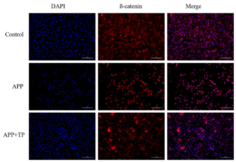
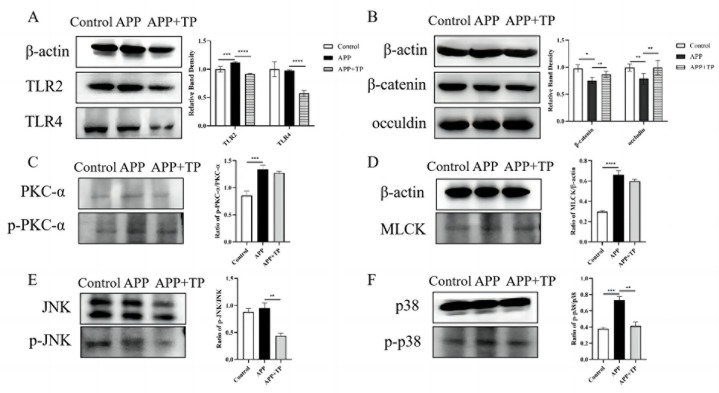
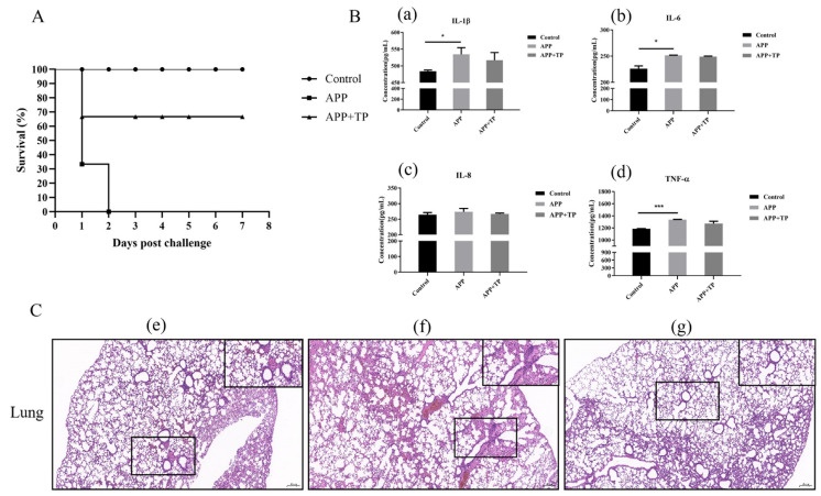
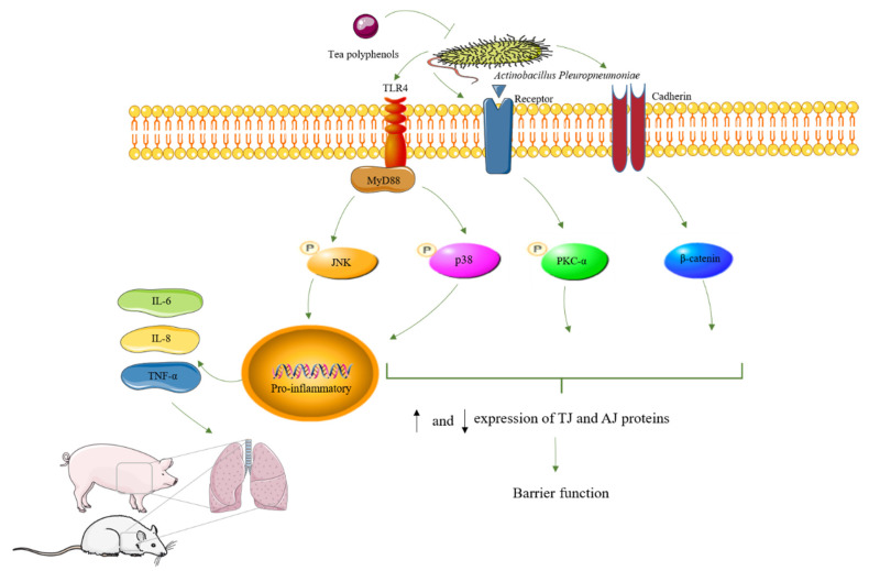
Actinobacillus pleuropneumoniae (APP) is the causative pathogen of porcine pleuropneumonia, a highly contagious respiratory disease in the pig industry. The increasingly severe antimicrobial resistance in APP urgently requires novel antibacterial alternatives for the treatment of APP infection. In this study, we investigated the effect of tea polyphenols (TP) against APP. MIC and MBC of TP showed significant inhibitory effects on bacteria growth and caused cellular damage to APP. Furthermore, TP decreased adherent activity of APP to the newborn pig tracheal epithelial cells (NPTr) and the destruction of the tight adherence junction proteins β-catenin and occludin. Moreover, TP improved the survival rate of APP infected mice but also attenuated the release of the inflammation-related cytokines IL-6, IL-8, and TNF-α. TP inhibited activation of the TLR/MAPK/PKC-MLCK signaling for down-regulated TLR-2, TLR4, p-JNK, p-p38, p-PKC-α, and MLCK in cells triggered by APP. Collectively, our data suggest that TP represents a promising therapeutic agent in the treatment of APP infection.

Keywords: Actinobacillus pleuropneumoniae; TLR-4/MAPK/PKC-MLCK signaling; epithelial barrier; tea polyphenols.


Conflict of interest statement

The authors declare no conflict of interest.

Figures

Figure 1
Figure 1
(A): Kinetics of the antimicrobial effects of TP on APP. APP and TP (0 MIC, 1/2 MIC, 1 MIC, and 2 MIC) were co-cultured for 0, 1, 2, 3, 4, and 5 h. The number of colonies was counted, and kinetics curves were constructed. (B): Transmission electron microscope and scanning electron microscopy analysis of APP. (a): Control untreated bacteria via TEM, the bar at the bottom right means 5.0 µm. High-magnification image of area indicated in upper right corner, the bar at the bottom right means 500 nm. (b): Bacteria treated with TP via TEM, the bar at the bottom right means 5.0 µm. High-magnification image of area indicated in upper right corner, the bar at the bottom right means 500 nm. (c): Untreated bacteria control analysis via SEM, the bar at the bottom right means 2.0 μm. (d): Bacteria treated with TP analysis via SEM, the bar at the bottom right means 2.0 μm. (C): The cell viability of NPTr cells of TP treatment. (D): The adherent ability of APP to NPTr cells or NPTr cells pretreatment with TP. Statistical analysis was performed by Student’s t-test. n = 3 in each group. Results are expressed as the mean ± SD of three independent experiments. * p < 0.05, *** p < 0.001.
Figure 2
Figure 2
(AC): Effect of APP on the Secretion of Inflammatory Factors in NPTr Cells or NPTr Cells Pretreated with TP. (D,E): The mRNA levels of TLR2 and TLR4 in NPTr cells and TP pretreatment cells infection with APP. Statistical analysis was performed by Student’s t-test. n = 3 in each group, ** p < 0.01, *** p < 0.001.
Figure 3
Figure 3
Immunofluorescence localization of occluding and β-catenin in NPTr cells. Scale bar indicated 100 μm.
Figure 3
Figure 3
Immunofluorescence localization of occluding and β-catenin in NPTr cells. Scale bar indicated 100 μm.
Figure 4
Figure 4
(A,B): The protein levels of TLR2, TLR4, ß-catenin, and occludin in NPTr cells and TP pretreatment cells infection with APP. GAPDH in whole cell lysates was detected as the loading control. (C): the ratio of p-PKC-α/PKC-α, (D): the ratio of MLCK: Effects of TP on PKC and MLCK pathways in NPTrs activated by APP. (E): the ratio of p-JNK/JNK, (F): the ratio of p-p38/p38: Effect of TP on phospho-JNK, and -p38 and total -JNK, and -p38 expression in NPTrs by Western blotting. (mean ± SD, n = 3).* p < 0.05, ** p < 0.01, *** p < 0.001, **** p < 0.0001.
Figure 5
Figure 5
(A): Survival curves for mice in infection experiment. (B): Secretion of IL-1ß, IL-6, IL-8, and TNF-α in the serum of mice infected with APP, as measured by ELISA. * p < 0.05, *** p < 0.001. (a): IL-1β production in serum of APP-infected mice affected by TP. (b): IL-6 production in serum of APP-infected mice affected by TP. (c): IL-8 production in serum of APP-infected mice affected by TP. (d): TNF-α production in serum of APP-infected mice afected by TP. (C): Histopathology of representative lung tissues from BALB/c mice and TP pretreatment mice infected with APP. (e): control group. (f): mice with APP infection group. (g): mice administered with TP. The black band at the bottom left of each picture indicates the scale bar (200 μm).
Figure 6
Figure 6
Schematic representation of TP in APP invasion of tracheal epithelial cells. “P” indicate phosphorylation. arrows (“↑” indicate increasing, “↓” indicate decreasing).

Similar articles

Cited by

References

    1. Caruana M., Vassallo N. Tea Polyphenols in Parkinson’s Disease. Adv. Exp. Med. Biol. 2015;863:117–137. - PubMed
    1. Guo Y.J., Sun L.Q., Yu B.Y., Qi J. An integrated antioxidant activity fingerprint for commercial teas based on their capacities to scavenge reactive oxygen species. Food Chem. 2017;237:645–653. doi: 10.1016/j.foodchem.2017.05.024. - DOI - PubMed
    1. Li Y., Gao X., Lou Y. Interactions of tea polyphenols with intestinal microbiota and their implication for cellular signal conditioning mechanism. J. Food Biochem. 2019;43:e12953. doi: 10.1111/jfbc.12953. - DOI - PubMed
    1. Zou L.-Q., Liu W., Liu W.-L., Liang R.-H., Li T., Liu C.-M., Cao Y.-L., Niu J., Liu Z. Characterization and Bioavailability of Tea Polyphenol Nanoliposome Prepared by Combining an Ethanol Injection Method with Dynamic High-Pressure Microfluidization. J. Agric. Food Chem. 2014;62:934–941. doi: 10.1021/jf402886s. - DOI - PubMed
    1. Xu X., Zhou X.D., Wu C.D. The tea catechin epigallocatechin gallate suppresses cariogenic virulence factors of Streptococcus mutans. Antimicrob. Agents Chemother. 2011;55:1229–1236. doi: 10.1128/AAC.01016-10. - DOI - PMC - PubMed