. 2023 Jun 28;13(7):1463.
doi: 10.3390/life13071463.

Phosphorylation Mimetic of Myosin Regulatory Light Chain Mitigates Cardiomyopathy-Induced Myofilament Impairment in Mouse Models of RCM and DCM

Affiliations

Phosphorylation Mimetic of Myosin Regulatory Light Chain Mitigates Cardiomyopathy-Induced Myofilament Impairment in Mouse Models of RCM and DCM

Katarzyna Kazmierczak et al. Life (Basel). .

Abstract

This study focuses on mimicking constitutive phosphorylation in the N-terminus of the myosin regulatory light chain (S15D-RLC) as a rescue strategy for mutation-induced cardiac dysfunction in transgenic (Tg) models of restrictive (RCM) and dilated (DCM) cardiomyopathy caused by mutations in essential (ELC, MYL3 gene) or regulatory (RLC, MYL2 gene) light chains of myosin. Phosphomimetic S15D-RLC was reconstituted in left ventricular papillary muscle (LVPM) fibers from two mouse models of cardiomyopathy, RCM-E143K ELC and DCM-D94A RLC, along with their corresponding Tg-ELC and Tg-RLC wild-type (WT) mice. The beneficial effects of S15D-RLC in rescuing cardiac function were manifested by the S15D-RLC-induced destabilization of the super-relaxed (SRX) state that was observed in both models of cardiomyopathy. S15D-RLC promoted a shift from the SRX state to the disordered relaxed (DRX) state, increasing the number of heads readily available to interact with actin and produce force. Additionally, S15D-RLC reconstituted with fibers demonstrated significantly higher maximal isometric force per cross-section of muscle compared with reconstitution with WT-RLC protein. The effects of the phosphomimetic S15D-RLC were compared with those observed for Omecamtiv Mecarbil (OM), a myosin activator shown to bind to the catalytic site of cardiac myosin and increase myocardial contractility. A similar SRX↔DRX equilibrium shift was observed in OM-treated fibers as in S15D-RLC-reconstituted preparations. Additionally, treatment with OM resulted in significantly higher maximal pCa 4 force per cross-section of muscle fibers in both cardiomyopathy models. Our results suggest that both treatments with S15D-RLC and OM may improve the function of myosin motors and cardiac muscle contraction in RCM-ELC and DCM-RLC mice.

Keywords: N-terminal protein modification; S15D-RLC phosphomimetic; Tg mice; cardiomyopathy; myosin ELC; myosin RLC; super-relaxed state (SRX).


Conflict of interest statement

The authors declare no conflict of interest. The funders had no role in the design of the study; in the collection, analyses, or interpretation of data; in the writing of the manuscript, or in the decision to publish the results.

Figures

Figure 1
Figure 1
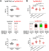
Force measurements in skinned LVPM from Tg-WT ELC (A) and Tg-E143K ELC (B) mice reconstituted with recombinant RLC proteins. Maximal force (top panels), Ca2+-sensitivity of force (middle panels), and force–pCa relationship (bottom panels) were measured in LVPM fibers from Tg-WT ELC and Tg-E143K mice. The fibers were depleted of endogenous RLC and reconstituted with recombinant WT-RLC (black-filled symbols) and S15D-RLC (green-filled symbols) proteins. Data are the average ± SD of n = 7–8 reconstituted LVPM fibers from Tg-WT ELC (2 animals-1F, 1M) and n = 9–10 reconstituted LVPM fibers from Tg-E143K ELC (3 animals-2F, 1M). Significance (p values) was calculated by Student’s t-test.
Figure 2
Figure 2
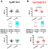
Summary of the SRX study for LVPM fibers from Tg-WT ELC (A) and Tg-E143K ELC (B) reconstituted with WT-RLC versus phosphomimetic S15D-RLC. The proportion of myosin heads in SRX state (top panels) and DRX states (bottom panels) is shown for native (clear points), WT- (black-filled points), and S15D- (green-filled points) RLC reconstituted Tg fibers. Note that % SRX is increased in native Tg-E143K fibers compared with Tg-WT ELC native controls (~71% versus ~61%). For Tg-E143K fibers reconstituted with WT-RLC or S15D-RLC, reconstitution with the latter significantly decreased % of myosin heads in SRX (~56%) compared with WT-RLC-reconstituted (68%) or native Tg-E143K fibers (71%). Data are expressed as mean ± SD for n = 14 Tg-WT ELC native fibers (4 animals-2F, 2M) and n = 6–7 (4 animals-2F, 2M) for Tg-WT ELC fibers reconstituted with WT-RLC or S15D-RLC. For Tg-E143K set, n = 13 fibers for native Tg-E143K (4 animals-2F, 2M) and n = 10–11 fibers for Tg-E143K (5 animals-3F, 2M) reconstituted with WT-RLC or S15D-RLC were used. The significance was calculated using one-way ANOVA with Tukey’s multiple comparison test.
Figure 3
Figure 3
Summary of the SRX study performed on reconstituted LVPM fibers from the model of DCM, Tg-D94A mice. (A) SRX comparison between Tg-WT RLC and Tg-D94A RLC mice. (B) The proportion of myosin heads in the SRX (top) and DRX (bottom) states. LVPM fibers reconstituted with WT-RLC are shown in black-filled symbols, while those reconstituted with S15D-RLC are shown in green-filled symbols. D94A-RLC-reconstituted fibers are depicted with red-filled symbols. Data are expressed as mean ± SD of n = N° fibers: n = 13, 4 animals-3F, 1M for native Tg-D94A, n = 17, 5 animals-2F, 3M for Tg-WT native, and n = 8–13, 4 animals-1F, 3M for LVPM fibers from Tg-D94A reconstituted with RLC proteins. The significance was calculated by Student’s t-test (A) and one-way ANOVA with Tukey’s multiple-comparison test (B).
Figure 4
Figure 4
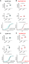
Contractile function in skinned LVPM fibers from Tg-WT ELC (A), Tg-E143K ELC (B), Tg-WT RLC (C), and Tg-D94A RLC (D) mice treated with 3 µM OM versus placebo (pCa 8 buffer). Maximal force (top panels), Ca2+-sensitivity of force (middle panels), and force–pCa relationships (bottom panels) were measured in fibers treated with 3 µM OM (cyan-filled points) compared with placebo (pCa 8 solution) (clear points). Data are the average ± SD of n = 4–8 fibers (1–2 mice) per group. p values were calculated by Student’s t-test.
Figure 5
Figure 5
Rescue of SRX↔DRX equilibrium in LVPM from RCM-ELC and DCM-RLC mouse models treated with 3 µM OM. Fibers treated with 3 µM OM (cyan-filled points) were compared with placebo-treated controls (clear points). (A) Tg-WT ELC, (B) Tg-E143K ELC, (C) Tg-WT RLC, and (D). Tg-D94A RLC mice. Data are the average ± SD of n = 9–14 fibers of 1–2 mice per group. p values were calculated by Student’s t-test.
Figure 5
Figure 5
Rescue of SRX↔DRX equilibrium in LVPM from RCM-ELC and DCM-RLC mouse models treated with 3 µM OM. Fibers treated with 3 µM OM (cyan-filled points) were compared with placebo-treated controls (clear points). (A) Tg-WT ELC, (B) Tg-E143K ELC, (C) Tg-WT RLC, and (D). Tg-D94A RLC mice. Data are the average ± SD of n = 9–14 fibers of 1–2 mice per group. p values were calculated by Student’s t-test.

References

    1. Geeves M.A., Holmes K.C. The molecular mechanism of muscle contraction. Adv. Protein Chem. 2005;71:161–193. - PubMed
    1. Tobacman L.S. Thin filament-mediated regulation of cardiac contraction. Annu. Rev. Physiol. 1996;58:447–481. doi: 10.1146/annurev.ph.58.030196.002311. - DOI - PubMed
    1. McKillop D.F., Geeves M.A. Regulation of the interaction between actin and myosin subfragment 1: Evidence for three states of the thin filament. Biophys. J. 1993;65:693–701. doi: 10.1016/S0006-3495(93)81110-X. - DOI - PMC - PubMed
    1. Brunello E., Fusi L., Ghisleni A., Park-Holohan S.J., Ovejero J.G., Narayanan T., Irving M. Myosin filament-based regulation of the dynamics of contraction in heart muscle. Proc. Natl. Acad. Sci. USA. 2020;117:8177–8186. doi: 10.1073/pnas.1920632117. - DOI - PMC - PubMed
    1. Wang Y., Yuan C.C., Kazmierczak K., Szczesna-Cordary D., Burghardt T.P. Single cardiac ventricular myosins are autonomous motors. Open Biol. 2018;8:170240. doi: 10.1098/rsob.170240. - DOI - PMC - PubMed