

Review
doi: 10.3390/medicina59071223.
Multimodality Cardiac Imaging in COVID-19 Infection
Affiliations
- PMID: 37512035
- PMCID: PMC10384118
- DOI: 10.3390/medicina59071223
Item in Clipboard
Review
Multimodality Cardiac Imaging in COVID-19 Infection
Medicina (Kaunas).
.
Abstract
COVID-19 infection often produces cardiovascular complications, which can range from mild to severe and influence the overall prognosis. Imaging is the cornerstone for diagnosing initial COVID-19 cardiovascular involvement as well as treatment guidance. In this review, we present the current state of the literature on this subject while also emphasizing possible algorithms for indicating and executing these investigations.
Keywords: COVID-19; cardiac magnetic resonance; echocardiography; multimodality imaging; myocarditis.
Conflict of interest statement
The authors declare no conflict of interest.
Figures

Echocardiographic markers of right ventricular impairment. (A) Significant tricuspid regurgitation. (B) High tricuspid gradient. (C) Right ventricular disfunction demonstrated by low TAPSE. (D) Short pulmonary acceleration time.

The Lake Louise criteria for diagnosis of myocarditis by cardiac magnetic resonance (reproduced after Ferreira et al.) [31].

Typical myocarditis as seen on the CMR of a 16-year-old patient with COVID-19 infection. (A). Inferior apical wall T2 hypersignal on a short-axis plane. (B) High native T2 on a four-chamber T2 mapping sequence. (C) High native T1 on a four-chamber T1 mapping sequence. (D) Inferior wall late enhancement on the intramyocardial and epicardial layers of the inferior wall on a PSIR short-axis plane.
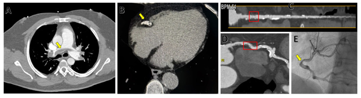
Examples of CT findings in COVID-19 patients. (A) Bilateral pulmonary artery embolism (yellow arrow) on an axial plane. (B) Large coronary calcified plaque on the right coronary artery (yellow arrow). (C) Straightened multiplanar reconstruction (MPR) of the right coronary artery showing moderate stenosis (60%–red box) in coronary CT angiography. (D) Curved multiplanar reconstruction (MPR) of the right coronary artery showing moderate stenosis (60%–red box) in coronary CT angiography. (E) Invasive coronary angiography confirming moderate stenosis (yellow arrow) of the right coronary artery. ((B–E) are for the same COVID-19 patient with chest pain, who also presented anomalous origin in the left anterior descending and circumflex arteries).

Imaging algorithm in confirmed COVID-19 patients with chest pain. SD, standard deviation; LV, left ventricle; RV, right ventricle; STEMI, ST-elevation myocardial infarction; CAD, coronary artery disease; PE, pulmonary embolism.
References
-
- Esposito A., Palmisano A., Cao R., Rancoita P., Landoni G., Grippaldi D., Boccia E., Cosenza M., Messina A., La Marca S., et al. Quantitative assessment of lung involvement on chest CT at admission: Impact on hypoxia and outcome in COVID-19 patients. Clin. Imaging. 2021;77:194. doi: 10.1016/j.clinimag.2021.04.033. - DOI - PMC - PubMed
-
- Lopes R.D., Macedo A.V.S., de Barros E Silva P.G.M., Moll-Bernardes R.J., Dos Santos T.M., Mazza L., Feldman A., D’Andréa Saba Arruda G., de Albuquerque D.C., Camiletti A.S., et al. Effect of Discontinuing vs Continuing Angiotensin-Converting Enzyme Inhibitors and Angiotensin II Receptor Blockers on Days Alive and Out of the Hospital in Patients Admitted With COVID-19: A Randomized Clinical Trial. JAMA. 2021;325:254. doi: 10.1001/jama.2020.25864. - DOI - PMC - PubMed
Publication types
MeSH terms
LinkOut - more resources
Full Text Sources
Medical
