Multicenter Study
. 2023 Jul 18;59(7):1323.
doi: 10.3390/medicina59071323.

Transcatheter Arterial Embolization (TAE) of Cancer-Related Bleeding

Affiliations
Multicenter Study

Transcatheter Arterial Embolization (TAE) of Cancer-Related Bleeding

Roberto Minici et al. Medicina (Kaunas). .

Abstract

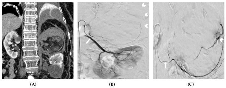
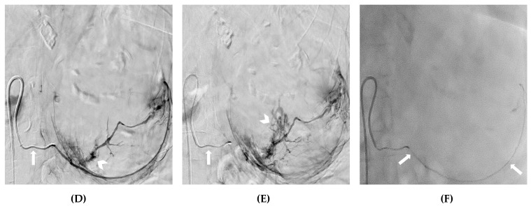
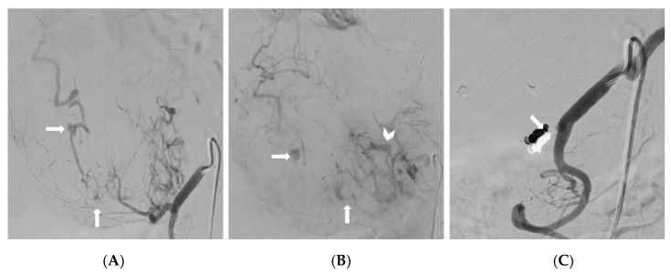
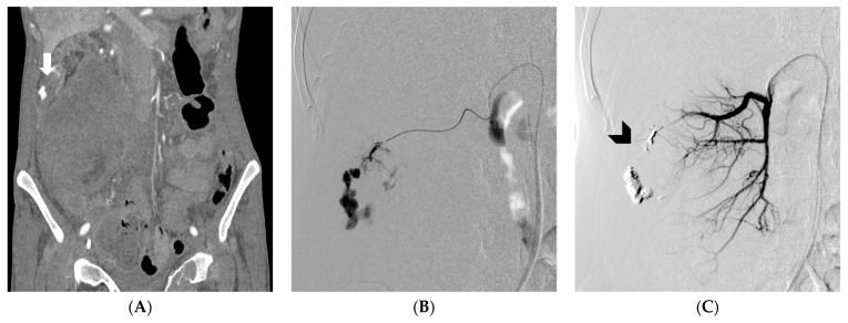
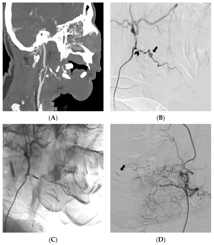
Background and Objectives: Roughly 10% of cancer patients experience an episode of bleeding. The bleeding severity can range from occasional trivial bleeds to major bleeding. The treatment for the bleeding may vary, depending on the clinical condition and anatomical site, and may include various strategies, among which TAE is a cornerstone of major bleeding management. However, the existing literature on tumor hemorrhages is inconsistent. The objective of this multicenter retrospective cohort study was to evaluate the effectiveness and safety of arterial embolization in the treatment of tumor hemorrhages in patients with solid cancers. Materials and Methods: The data for patients with solid cancers undergoing TAE for the management of tumor hemorrhages from January 2020 to May 2023 were gathered. Results: A total of 92 patients with cancer-related bleeding were treated between January 2020 and May 2023. No bleeding was detected by X-ray angiography (XA) in 12 (13%) cases; therefore, a blind embolization was performed. The most common bleeding site was the liver (21.7%). A total of 66 tumor hemorrhages were spontaneous. The most commonly used embolic agent was polyvinyl alcohol (PVA) particles (30.4%). Technical success was achieved in 82 (89.1%) cases, with an 84.8% clinical success rate related to 14 cases of rebleeding. Proximal embolization was performed for 19 (20.7%) patients. Complications were recorded for 10 (10.9%) patients. The 30-day bleeding-related mortality was 15.2%. The technical success, clinical success, proximal embolization rate, and 30-day rebleeding were worse in the subset of patients undergoing TAE with coils. Conclusions: Transcatheter arterial embolization (TAE) represents a viable and potentially life-saving therapeutic approach in the management of tumor hemorrhages, demonstrating a notable effectiveness and safety. The TAE of bleeding tumors using coils resulted in a higher rate of non-superselective proximal embolization, with a trend toward lower clinical success rates and higher rebleeding episodes.

Keywords: TAE; bleeding; cancer; embolic agents; embolization; endovascular; hemorrhage; tumor.


Conflict of interest statement

The authors declare no conflict of interest.

Figures

Figure 1
Figure 1
(A) A CTA showing spontaneous retroperitoneal bleeding due to an unknown, ruptured, adrenal myelolipoma. (B) A digital subtraction angiography depicting the inferior adrenal artery arising (arrow) from the proximal left renal artery, along with the shadow (arrowheads) of the ruptured myelolipoma displacing the left kidney downwards. (C,D,E) The superselective catheterization of the target artery with a microcatheter (arrow) and digital subtraction angiography, showing a massive intratumoral hemorrhage (arrowheads). (F) A fluoroscopy demonstrating the embolization of the inferior adrenal artery, using PVA particles mixed with iodinated contrast media (arrows).
Figure 1
Figure 1
(A) A CTA showing spontaneous retroperitoneal bleeding due to an unknown, ruptured, adrenal myelolipoma. (B) A digital subtraction angiography depicting the inferior adrenal artery arising (arrow) from the proximal left renal artery, along with the shadow (arrowheads) of the ruptured myelolipoma displacing the left kidney downwards. (C,D,E) The superselective catheterization of the target artery with a microcatheter (arrow) and digital subtraction angiography, showing a massive intratumoral hemorrhage (arrowheads). (F) A fluoroscopy demonstrating the embolization of the inferior adrenal artery, using PVA particles mixed with iodinated contrast media (arrows).
Figure 2
Figure 2
(A,B) The selective catheterization of the common hepatic artery, and the subsequent digital subtraction angiography, demonstrating a large ruptured hepatocellular carcinoma with multiple pseudoaneurysms (arrows) and arteriovenous shunt (arrowhead); the origin of the right hepatic artery from the superior mesenteric artery noted at the CT angiography was also confirmed. (C) A digital subtraction angiography depicting an effective embolization of the target artery with EVOH copolymer; the onyx cast can be noted (arrow).
Figure 3
Figure 3
(A) demonstrates a CT angiography revealing spontaneous retroperitoneal bleeding caused by a ruptured pseudoaneurysm (arrow), originating from a renal tumor. (B) displays a digital subtraction angiography confirming the presence of a ruptured pseudoaneurysm, arising from a feeding artery of the tumor. (C) exhibits DSA, illustrating successful embolization, using an EVOH copolymer cast (arrowhead) (from [19], by MDPI, Basel, Switzerland, licensed under CC BY 4.0).
Figure 4
Figure 4
In this case of locally advanced squamous cell carcinoma of the tongue, (A) a CT angiography with maximum intensity projection reformatted in the sagittal plane, and (B) a digital subtraction angiography of the external carotid artery, were conducted. (A,B) Significant oral bleeding originated from a pseudoaneurysm (arrow) located in the context of an irregular arterial wall profile (arrowhead). (C) The successful embolization of the lingual artery was achieved using an EVOH copolymer, as demonstrated in the angiogram. (D) A contralateral digital subtraction angiography excluded retrograde bleeding, and the presence of the onyx cast can be observed (arrow) (from [19] by MDPI, Basel, Switzerland, licensed under CC BY 4.0).

References

    1. Pereira J., Phan T. Management of bleeding in patients with advanced cancer. Oncologist. 2004;9:561–570. doi: 10.1634/theoncologist.9-5-561. - DOI - PubMed
    1. Johnstone C., Rich S.E. Bleeding in cancer patients and its treatment: A review. Ann. Palliat. Med. 2018;7:265–273. doi: 10.21037/apm.2017.11.01. - DOI - PubMed
    1. Schulman S., Kearon C., Subcommittee on Control of Anticoagulation of the Scientific and Standardization Committee of the International Society on Thrombosis and Haemostasis Definition of major bleeding in clinical investigations of antihemostatic medicinal products in non-surgical patients. J. Thromb. Haemost. 2005;3:692–694. doi: 10.1111/j.1538-7836.2005.01204.x. - DOI - PubMed
    1. European Medicines Agency Clinical Investigation of Medicinal Products for the Prophylaxis of Venous Thromboembolic Risk in Non-Surgical Patients—Scientific Guideline. [(accessed on 20 May 2023)]. Available online: https://www.ema.europa.eu/en/clinical-investigation-medicinal-products-p....
    1. Cihoric N., Crowe S., Eychmüller S., Aebersold D.M., Ghadjar P. Clinically significant bleeding in incurable cancer patients: Effectiveness of hemostatic radiotherapy. Radiat. Oncol. 2012;7:132. doi: 10.1186/1748-717X-7-132. - DOI - PMC - PubMed

Publication types