. 2023 Jul 6;11(7):1762.
doi: 10.3390/microorganisms11071762.

Extracts from Wallis Sponges Inhibit Vibrio harveyi Biofilm Formation

Affiliations

Extracts from Wallis Sponges Inhibit Vibrio harveyi Biofilm Formation

Flore Caudal et al. Microorganisms. .

Abstract

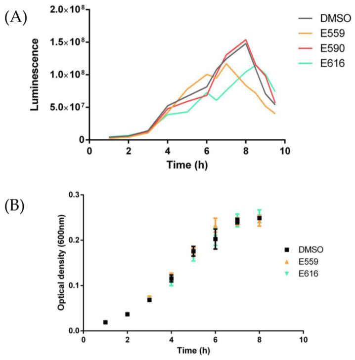
Pathogenic bacteria and their biofilms are involved in many human and animal diseases and are a major public health problem with, among other things, the development of antibiotic resistance. These biofilms are known to induce chronic infections for which classical treatments using antibiotic therapy are often ineffective. Sponges are sessile filter-feeding marine organisms known for their dynamic symbiotic partnerships with diverse microorganisms and their production of numerous metabolites of interest. In this study, we investigated the antibiofilm efficacy of different extracts from sponges, isolated in Wallis, without biocidal activity. Out of the 47 tested extracts, from 28 different genera, 11 showed a strong activity against Vibrio harveyi biofilm formation. Moreover, one of these extracts also inhibited two quorum-sensing pathways of V. harveyi.

Keywords: Vibrio harveyi; anti-biofilm activity; biofilm; marine natural products; sponge extract.


Conflict of interest statement

The authors declare no conflict of interest. The funders had no role in the design of the study; in the collection, analyses, or interpretation of data; in the writing of the manuscript; or in the decision to publish the results.

Figures

Figure A1
Figure A1
Inhibition of P. aeruginosa MUC-N1 biofilm formation by different sponge extracts in microtiter plates. P. aeruginosa MUC-N1 was inoculated with sponge extracts (10 µg/mL) in 96-well microplates and incubated at 37 °C for 24 h to form biofilms. Sponge extracts have been grouped by genus. Bars represent means ± standard error of the mean for three replicates, and the dotted line corresponds to the DMSO control.
Figure A2
Figure A2
Inhibition of T. maritimum biofilm formation by different sponge extracts in microtiter plates. T. maritimum was inoculated with sponge extracts (10 µg/mL) in 96-well microplates and incubated at 28 °C for 24 h to form biofilms. Sponge extracts have been grouped by genus. Bars represent means ± standard error of the mean for three replicates, and the dotted line corresponds to the DMSO control.
Figure 1
Figure 1
Inhibition of V. harveyi ORM4 biofilm formation by different sponge extracts in microtiter plates. V. harveyi ORM4 was inoculated with SE (10 µg/mL) in 96-well microplates and incubated at 20 °C for 48 h to form biofilms. The dotted lines delimitate four groups of SEs, which have been distinguished according to their activity levels. The sponge genera are indicated. Bars represent means ± standard error of the mean for three replicates and the 100% dotted line corresponds to the DMSO SE-free control. * p < 0.05.
Figure 2
Figure 2
Percentage of glass surface covered by bacteria in the presence of sponge extracts compared to the SE-free control. V. harveyi ORM4 was inoculated with sponge extracts (10 µg/mL) in flow-cell chambers and bacteria were allowed to attach at 20 °C for 2 h. The sponge genera are indicated. Bars represent means ± standard error of the mean for three replicates and the dotted line corresponds to the DMSO control.
Figure 3
Figure 3
Light microscopic observation of the effect of nine sponge extracts on the adhesion of V. harveyi ORM4. V. harveyi ORM4 was inoculated with sponge extracts (10 µg/mL) in flow-cell chambers and allowed to attach on a glass surface at 20 °C for 2 h prior to observe them at 40× magnification. Images shown are representative of four images per condition from three independent experiments.
Figure 4
Figure 4
V. harveyi ORM4 biofilms formed in flow-cell chambers after incubation with different sponge extracts during the attachment step. V. harveyi ORM4 was inoculated with SEs (10 µg/mL) in flow cell chambers and biofilms were then grown at 20 °C for 24 h. Confocal laser microscopy observations with or without the addition of extracts are in 3D: top and side views are shown here.
Figure 5
Figure 5
Effect of sponge extracts on V. harveyi BB120 quorum sensing. (A) Luminescence of V. harveyi BB120 in the presence of 10 µg/mL extract or with DMSO as control. Luminescence is counted in relative light units. (B) Kinetic growth of V. harveyi BB120 in the presence of 10 µg/mL extract or SE-free DMSO as control; growth is shown as optical density at 600 nm.
Figure 6
Figure 6
Effect of sponge extracts on V. harveyi JAF375 (A), JMH597 (B), and JMH612 (C) quorum sensing. Luminescence of V. harveyi JAF375 (A), JMH597 (B), and JMH612 (C) in the presence of 10 µg/mL extract or with DMSO as control. Luminescence is shown as RLU.

Similar articles

Cited by

References

    1. Murray C.J., Ikuta K.S., Sharara F., Swetschinski L., Robles Aguilar G., Gray A., Han C., Bisignano C., Rao P., Wool E., et al. Global Burden of Bacterial Antimicrobial Resistance in 2019: A Systematic Analysis. Lancet. 2022;399:629–655. doi: 10.1016/S0140-6736(21)02724-0. - DOI - PMC - PubMed
    1. Yin W., Wang Y., Liu L., He J. Biofilms: The Microbial “Protective Clothing” in Extreme Environments. Int. J. Mol. Sci. 2019;20:3423. doi: 10.3390/ijms20143423. - DOI - PMC - PubMed
    1. Stewart P.S. Mechanisms of Antibiotic Resistance in Bacterial Biofilms. Int. J. Med. Microbiol. IJMM. 2002;292:107–113. doi: 10.1078/1438-4221-00196. - DOI - PubMed
    1. Funari R., Shen A.Q. Detection and Characterization of Bacterial Biofilms and Biofilm-Based Sensors. ACS Sens. 2022;7:347–357. doi: 10.1021/acssensors.1c02722. - DOI - PubMed
    1. Anderl J.N., Franklin M.J., Stewart P.S. Role of Antibiotic Penetration Limitation in Klebsiella Pneumoniae Biofilm Resistance to Ampicillin and Ciprofloxacin. Antimicrob. Agents Chemother. 2000;44:1818–1824. doi: 10.1128/AAC.44.7.1818-1824.2000. - DOI - PMC - PubMed

LinkOut - more resources