. 2023 Jul 11;11(7):1789.
doi: 10.3390/microorganisms11071789.

A Severe Acute Respiratory Syndrome Coronavirus 2 Anti-Spike Immunoglobulin G Assay: A Robust Method for Evaluation of Vaccine Immunogenicity Using an Established Correlate of Protection

Affiliations

A Severe Acute Respiratory Syndrome Coronavirus 2 Anti-Spike Immunoglobulin G Assay: A Robust Method for Evaluation of Vaccine Immunogenicity Using an Established Correlate of Protection

Mingzhu Zhu et al. Microorganisms. .

Abstract

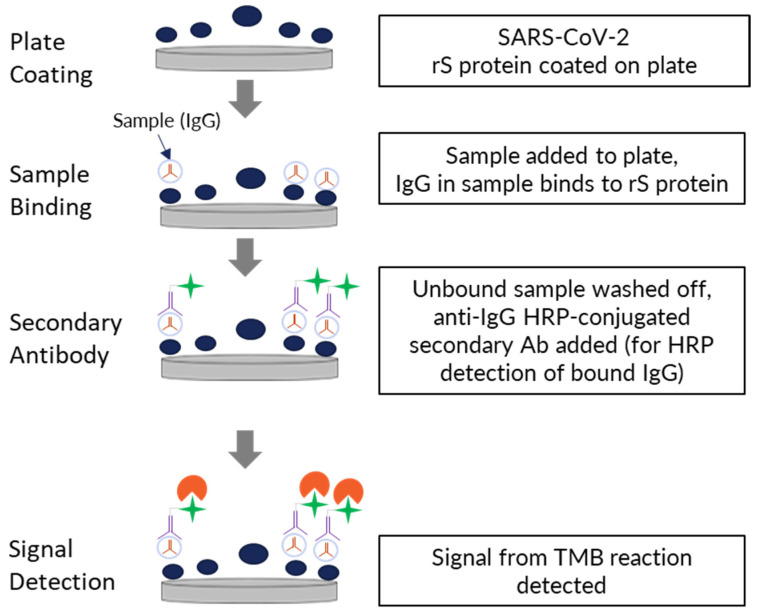
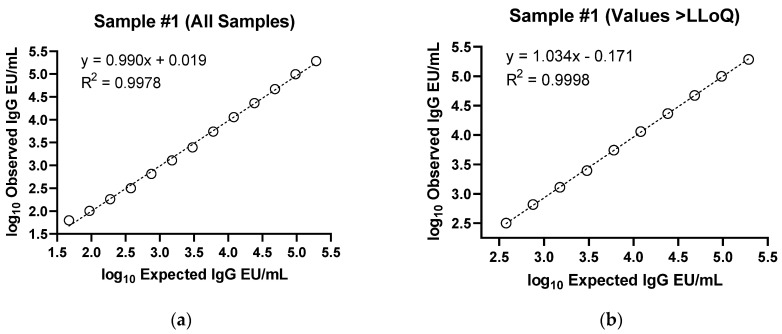
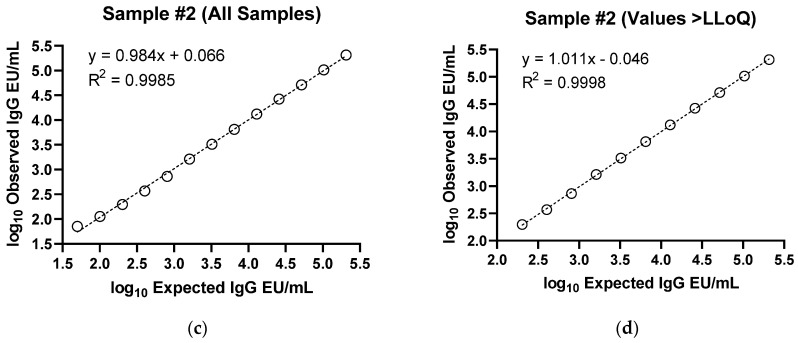
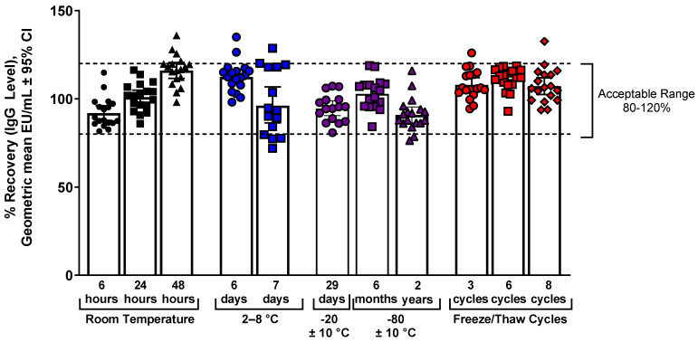
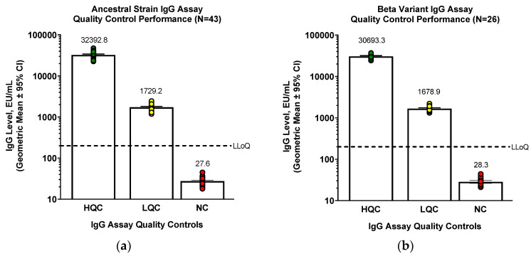
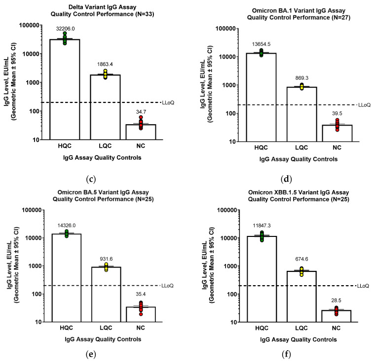
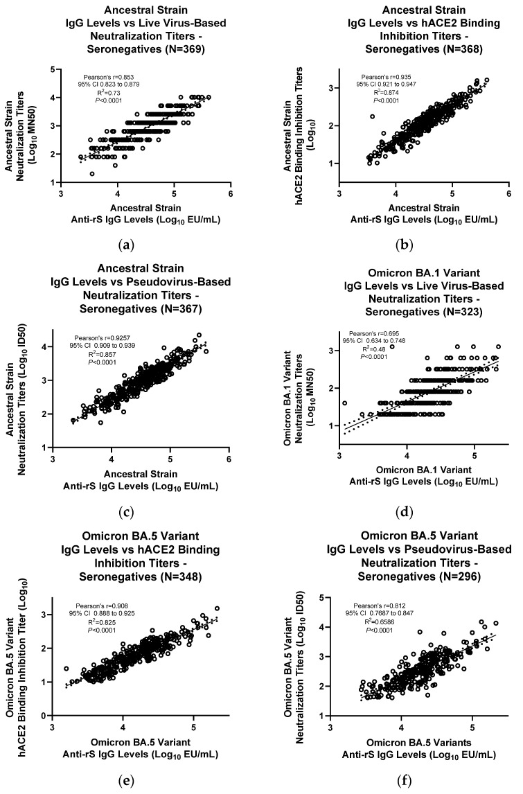
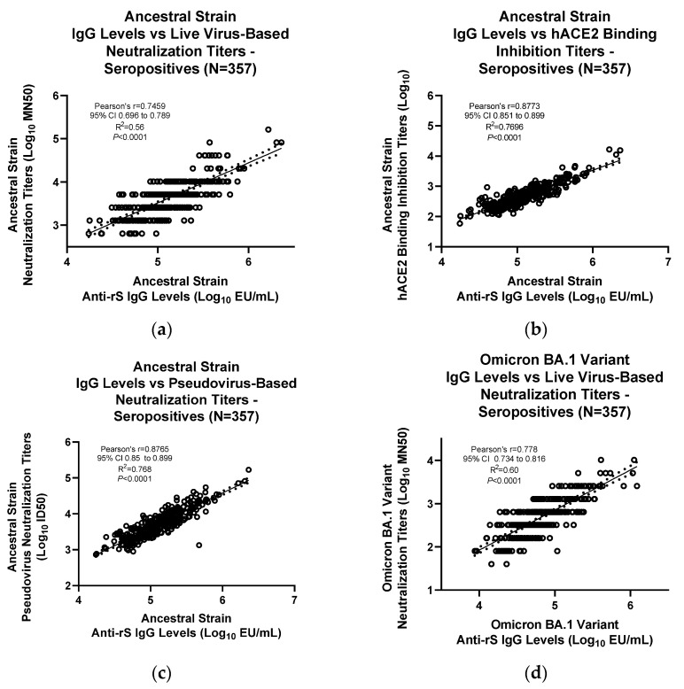
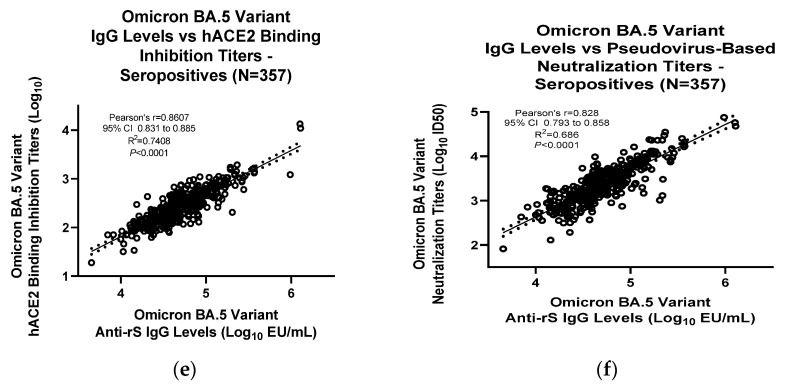
As the COVID-19 pandemic continues, variants of severe acute respiratory syndrome coronavirus 2 (SARS-CoV-2) continue to emerge. Immunogenicity evaluation of vaccines and identification of correlates of protection for vaccine effectiveness is critical to aid the development of vaccines against emerging variants. Anti-recombinant spike (rS) protein immunoglobulin G (IgG) quantitation in the systemic circulation (serum/plasma) is shown to correlate with vaccine efficacy. Thus, an enzyme-linked immunosorbent assay (ELISA)-based binding assay to detect SARS-CoV-2 (ancestral and variant strains) anti-rS IgG in human serum samples was developed and validated. This assay successfully met acceptance criteria for inter/intra-assay precision, specificity, selectivity, linearity, lower/upper limits of quantitation, matrix effects, and assay robustness. The analyte in serum was stable for up to 8 freeze/thaw cycles and 2 years in -80 °C storage. Similar results were observed for the Beta, Delta, and Omicron BA.1/BA.5/XBB.1.5 variant-adapted assays. Anti-rS IgG assay results correlated significantly with neutralization and receptor binding inhibition assays. In addition, usage of international reference standards allows data extrapolation to WHO international units (BAU/mL), facilitating comparison of results with other IgG assays. This anti-rS IgG assay is a robust, high-throughput method to evaluate binding IgG responses to S protein in serum, enabling rapid development of effective vaccines against emerging COVID-19 variants.

Keywords: Binding antibodies; COVID-19; ELISA; IgG; Omicron BA.1/BA.5/XBB.1.5 SARS-CoV-2 variants; antibodies; correlate of protection; variants of concern.


Conflict of interest statement

All the authors are employees and stockholders of Novavax, Inc.

Figures

Figure 1
Figure 1
Procedure for performing the anti-rS IgG detection assay. Ab—antibody; IgG—immunoglobulin G; HRP—horseradish peroxidase; rS—recombinant spike; SARS-CoV-2—severe acute respiratory syndrome coronavirus 2; TMB—3,3′5,5′-tetramethylbenzidine.
Figure 2
Figure 2
IgG assay ancestral strain linearity. Two samples positive for anti-rS IgG were diluted in 1:2 series and tested in 6 assay runs. Linearity was then evaluated by calculating the precision and accuracy of the concentration at each dilution and the slope of linear regression lines for each sample. For both samples, dilutions are shown from undiluted to 1:512 dilution. (a) Sample #1 with all samples shown, (b) Sample #1 with only values above LLoQ (200 EU/mL), (c) Sample #2 with all samples shown, and (d) Sample #2 with only values above LLoQ are presented.
Figure 2
Figure 2
IgG assay ancestral strain linearity. Two samples positive for anti-rS IgG were diluted in 1:2 series and tested in 6 assay runs. Linearity was then evaluated by calculating the precision and accuracy of the concentration at each dilution and the slope of linear regression lines for each sample. For both samples, dilutions are shown from undiluted to 1:512 dilution. (a) Sample #1 with all samples shown, (b) Sample #1 with only values above LLoQ (200 EU/mL), (c) Sample #2 with all samples shown, and (d) Sample #2 with only values above LLoQ are presented.
Figure 3
Figure 3
IgG assay ancestral strain temperature and freeze/thaw stability. Samples were stored at different temperature conditions or were subjected to multiple freeze/thaw cycles and then used in the IgG assay. The acceptable recovery range was 80% to 120% of the reference condition values (dashed lines).
Figure 4
Figure 4
IgG assay ancestral strain and variants quality control standards performance. (a) For ancestral strain, (b) Beta variant, (c) Delta variant, (d) Omicron BA.1 variant, (e) Omicron BA.5 variant, or (f) Omicron XBB.1.5 variant, QC samples were tested in the IgG detection assay to examine assay performance. One value was identified as an outlier for the NC group of XBB.1.5 and was removed from the analysis.
Figure 4
Figure 4
IgG assay ancestral strain and variants quality control standards performance. (a) For ancestral strain, (b) Beta variant, (c) Delta variant, (d) Omicron BA.1 variant, (e) Omicron BA.5 variant, or (f) Omicron XBB.1.5 variant, QC samples were tested in the IgG detection assay to examine assay performance. One value was identified as an outlier for the NC group of XBB.1.5 and was removed from the analysis.
Figure 5
Figure 5
Correlation of IgG assay results with other immunogenicity assays in SARS-CoV-2 seronegative serum samples. Results from the IgG assay analyses were correlated with results from other assays for (ac) ancestral strain, (d) Omicron BA.1 variant, or (e,f) Omicron BA.5 variant. For (c,f), a pseudovirus neutralization assay was performed as previously described [14]. For (a,d), a live wild-type virus neutralization assay was performed as previously described [13]. For (b,e), an hACE2 binding inhibition assay was performed as previously described [15]. Linear regression analysis was performed to compare results from the IgG assay and the other assays. ID50—median infectious dose; LOD—limit of detection; Log10—logarithm with base 10; MN50—50% microneutralization.
Figure 6
Figure 6
Correlation of IgG assay results with other immunogenicity assays in SARS-CoV-2 seropositive serum samples. Results from the IgG assay analyses were correlated with results from other assays for (ac) ancestral strain, (d) Omicron BA.1 variant, or (e,f) Omicron BA.5 variant. For (c,f), a pseudovirus neutralization assay was performed as previously described [14]. For (a,d), a live wild-type virus neutralization assay was performed as previously described [13]. For (b,e), an hACE2 binding inhibition assay was performed as previously described [15]. Linear regression analysis was performed to compare results from the IgG assay and the other assays. ID50—median infectious dose; LOD—limit of detection; Log10—logarithm with base 10; MN50—50% microneutralization.
Figure 6
Figure 6
Correlation of IgG assay results with other immunogenicity assays in SARS-CoV-2 seropositive serum samples. Results from the IgG assay analyses were correlated with results from other assays for (ac) ancestral strain, (d) Omicron BA.1 variant, or (e,f) Omicron BA.5 variant. For (c,f), a pseudovirus neutralization assay was performed as previously described [14]. For (a,d), a live wild-type virus neutralization assay was performed as previously described [13]. For (b,e), an hACE2 binding inhibition assay was performed as previously described [15]. Linear regression analysis was performed to compare results from the IgG assay and the other assays. ID50—median infectious dose; LOD—limit of detection; Log10—logarithm with base 10; MN50—50% microneutralization.

Similar articles

Cited by

References

    1. Centers for Disease Control and Prevention David J. Spencer CDC Museum—COVID-19 Timeline. [(accessed on 3 February 2023)]; Available online: https://www.cdc.gov/museum/timeline/covid19.html.
    1. Fendler A., de Vries E.G.E., GeurtsvanKessel C.H., Haanen J.B., Wörmann B., Turajlic S., von Lilienfeld-Toal M. COVID-19 vaccines in patients with cancer: Immunogenicity, efficacy and safety. Nat. Rev. Clin. Oncol. 2022;19:385–401. doi: 10.1038/s41571-022-00610-8. - DOI - PMC - PubMed
    1. Fiolet T., Kherabi Y., MacDonald C.J., Ghosn J., Peiffer-Smadja N. Comparing COVID-19 vaccines for their characteristics, efficacy and effectiveness against SARS-CoV-2 and variants of concern: A narrative review. Clin. Microbiol. Infect. 2022;28:202–221. doi: 10.1016/j.cmi.2021.10.005. - DOI - PMC - PubMed
    1. Liu Y., Rocklöv J. The effective reproductive number of the Omicron variant of SARS-CoV-2 is several times relative to Delta. J. Travel Med. 2022;29:taac037. doi: 10.1093/jtm/taac037. - DOI - PMC - PubMed
    1. Van Tilbeurgh M., Lemdani K., Beignon A.-S., Chapon C., Tchitchek N., Cheraitia L., Marcos Lopez E., Pascal Q., Le Grand R., Maisonnasse P., et al. Predictive Markers of Immunogenicity and Efficacy for Human Vaccines. Vaccines. 2021;9:579. doi: 10.3390/vaccines9060579. - DOI - PMC - PubMed