. 2023 Jul 20;11(7):1842.
doi: 10.3390/microorganisms11071842.

Lichenysin-like Polypeptide Production by Bacillus licheniformis B3-15 and Its Antiadhesive and Antibiofilm Properties

Affiliations

Lichenysin-like Polypeptide Production by Bacillus licheniformis B3-15 and Its Antiadhesive and Antibiofilm Properties

Vincenzo Zammuto et al. Microorganisms. .

Abstract

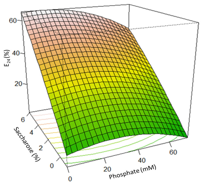
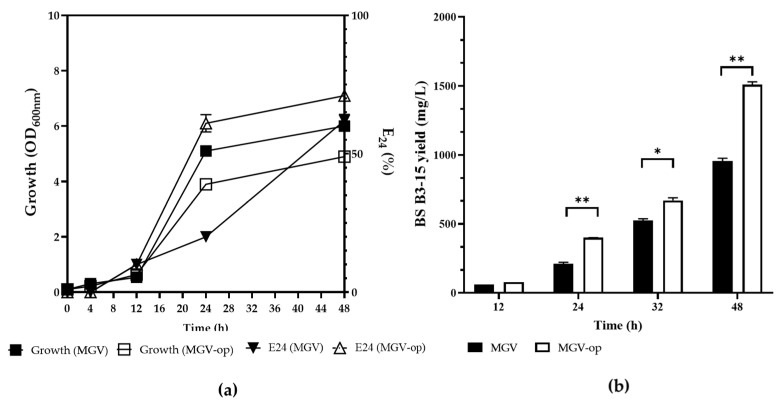
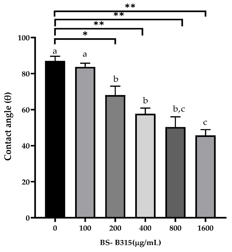
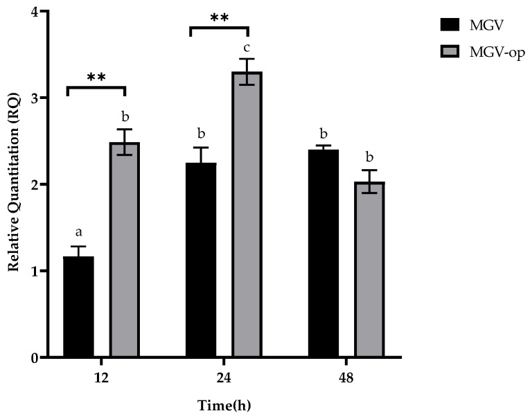
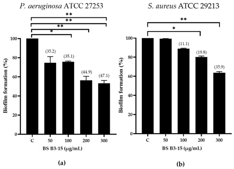
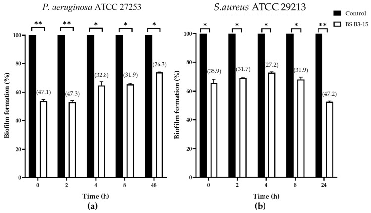
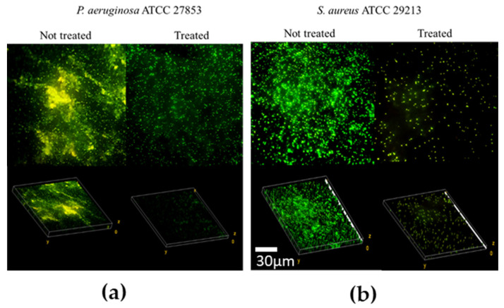
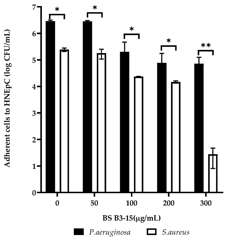
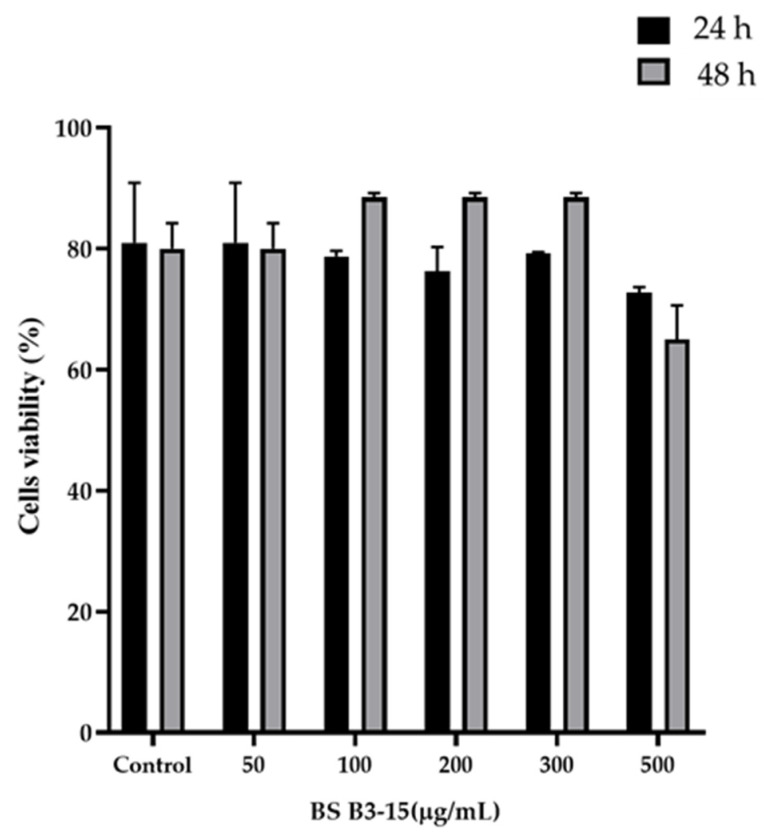
We report the ability of the crude biosurfactant (BS B3-15), produced by the marine, thermotolerant Bacillus licheniformis B3-15, to hinder the adhesion and biofilm formation of Pseudomonas aeruginosa ATCC 27853 and Staphylococcus aureus ATCC 29213 to polystyrene and human cells. First, we attempted to increase the BS yield, optimizing the culture conditions, and evaluated the surface-active properties of cell-free supernatants. Under phosphate deprivation (0.06 mM) and 5% saccharose, the yield of BS (1.5 g/L) increased by 37%, which could be explained by the earlier (12 h) increase in lchAA expression compared to the non-optimized condition (48 h). Without exerting any anti-bacterial activity, BS (300 µg/mL) prevented the adhesion of P. aeruginosa and S. aureus to polystyrene (47% and 36%, respectively) and disrupted the preformed biofilms, being more efficient against S. aureus (47%) than P. aeruginosa (26%). When added to human cells, the BS reduced the adhesion of P. aeruginosa and S. aureus (10× and 100,000× CFU/mL, respectively) without altering the epithelial cells' viability. As it is not cytotoxic, BS B3-15 could be useful to prevent or remove bacterial biofilms in several medical and non-medical applications.

Keywords: Bacillus; antiadhesive; antibiofilm; bioemulsifier; biosurfactant; human cell viability.


Conflict of interest statement

The authors declare no conflict of interest.

Figures

Figure 1
Figure 1
Effect of the saccharose (%) and phosphate (mM) concentrations on the emulsifying activity, expressed as E24 (%) of the B3-15 cell-free supernatant. The different colors on the surface plot represent the gradient range, from the lowest (green) to the greatest (light-red), for the BS production.
Figure 2
Figure 2
Comparison of the B. licheniformis B3-15 (a) growth (OD600nm) and the emulsifying index (E24%) of cell-free supernatants (1:1 kerosene) and (b) the crude biosurfactant yield (mg/L) at up to 48 h of incubation in non-optimized (MGV) and optimized media (MGV-op). Data are expressed as averages and standard deviations (n = 3). * Significantly different (p ≤ 0.05) or ** p ≤ 0.01.
Figure 3
Figure 3
ATR-FTIR spectrum of the crude BS B3-15 produced by B. licheniformis B3-15 in optimized conditions (MGV-op) after 48 h incubation at 45 °C.
Figure 4
Figure 4
Surface properties, measured as contact angle (θ) values, with or without the BS B3-15 at different concentrations (0, 100, 200, 400, 800, and 1600 µg/mL). The bars represent the data mean ± SD for three replicates (n = 3). Statistical differences were evaluated using two-way ANOVA with Tukey’s multiple comparisons test. Significantly different, * p ≤ 0.05 and ** p ≤ 0.01, compared with untreated controls. Different lowercase letters above the bar graph indicate significant statistical differences (p < 0.01).
Figure 5
Figure 5
Relative quantitation (RQ) of lchAA at different incubation times (12, 24, and 48 h) of B3-15 in MGV or MGV-op. The bars represent mean ± SD for three replicates (n = 3). Statistical differences were evaluated using two-way ANOVA with Tukey’s multiple comparisons tests. Significantly different ** p ≤ 0.01 compared with untreated controls. Different lowercase letters above the bar graph indicate significant statistical differences (p < 0.01).
Figure 6
Figure 6
Pseudomonas aeruginosa ATCC 27853 (a) and Staphylococcus aureus ATCC 29213 (b) biofilm formation (%) on polystyrene microplate without (C) or with the BS B3-15 at different concentrations (50, 100, 200, and 300 µg/mL). The bars represent the mean ± standard deviation for six replicates (n = 6). * p ≤ 0.05 and ** p ≤ 0.01 show significant differences compared with untreated controls. Data on biofilm reduction (%) are reported in brackets.
Figure 7
Figure 7
Pseudomonas aeruginosa ATCC 27853 (a) and Staphylococcus aureus ATCC 29213 (b) biofilm formation (%) on polystyrene microplate without (Control) or after the addition of the BS B3-15 (300 μg/mL) at different bacterial growth times (T0, T2, T4, T8) and after T48 for P. aeruginosa and T24 for S. aureus, when the biofilms were completely established. The bars represent mean ± SD for six replicates (n = 6) * p ≤ 0.05 and ** p ≤ 0.01 show significant differences compared with untreated controls. Data on biofilm reduction (%) are reported in brackets.
Figure 8
Figure 8
Confocal laser images (×600) of preformed biofilms from (a) Pseudomonas aeruginosa ATCC 27853 and (b) Staphylococcus aureus ATCC 29213 on polystyrene surfaces, not treated or treated with BS B3-15 (300 μg/mL).
Figure 9
Figure 9
Adherence of Pseudomonas aeruginosa ATCC 27853 and Staphylococcus aureus ATCC 29213 to human nasal epithelial cells (HNEpCs), expressed on a logarithmic scale of CFU/mL, in the presence of BS B3-15, after 2 h of incubation at 37 °C. The bars represent the mean ± standard deviation for three replicates (n = 3). Statistical differences were evaluated using ANOVA coupled with two-way Tukey’s multiple-comparison tests. * p ≤ 0.05 and ** p ≤ 0.01: significant differences compared with untreated controls.
Figure 10
Figure 10
Viability of HNEpC exposed to different BS B3-15 concentrations (from 50 to 500 µg/mL) after 24 h or 48 h. Bars represent the data’s mean ± SD for six replicates (n = 6).

References

    1. Kaczorek E., Pachholak A., Zdarta A., Smulek W. The impact of biosurfactant on microbial properties leading to hydrocarbon bioavailability. Colloids Interfaces. 2018;2:35. doi: 10.3390/colloids2030035. - DOI
    1. Janek T., Lukaszewicz M., Krasowska A. Identification and characterization of biosurfactants produced by the Arctic bacterium Pseudomonas putida BD2. Colloids Surf. B Biointerfaces. 2013;110:379–386. doi: 10.1016/j.colsurfb.2013.05.008. - DOI - PubMed
    1. De França Í.W.L., Lima A.P., Lemos J.A.M., Lemos C.G.F., Melo V.M.M., de Sant’ana H.B., Gonçalves L.R.B. Production of a biosurfactant by Bacillus subtilis ICA56 aiming bioremediation of impacted soils. Catal. Today. 2015;255:10–15. doi: 10.1016/j.cattod.2015.01.046. - DOI
    1. Perfumo A., Rancich I., Banat I.M. Possibilities and challenges for biosurfactants use in petroleum industry. In: Sen R., editor. Biosurfactants. Advances in Experimental Medicine and Biology. Springer; New York, NY, USA: 2010. pp. 135–145. - PubMed
    1. Smyth T.J.P., Perfumo A., Marchant R., Banat I.M. Isolation and analysis of low molecular weight microbial glycolipids. In: Timmis K.N., editor. Handbook of Hydrocarbon and Lipid Microbiology. Springer; Berlin, Germany: 2010. pp. 3705–3723.

LinkOut - more resources