. 2023 Jul 10;13(14):2040.
doi: 10.3390/nano13142040.

Incorporation/Enrichment of 3D Bioprinted Constructs by Biomimetic Nanoparticles: Tuning Printability and Cell Behavior in Bone Models

Affiliations

Incorporation/Enrichment of 3D Bioprinted Constructs by Biomimetic Nanoparticles: Tuning Printability and Cell Behavior in Bone Models

Tiziana Fischetti et al. Nanomaterials (Basel). .

Abstract

Reproducing in vitro a model of the bone microenvironment is a current need. Preclinical in vitro screening, drug discovery, as well as pathophysiology studies may benefit from in vitro three-dimensional (3D) bone models, which permit high-throughput screening, low costs, and high reproducibility, overcoming the limitations of the conventional two-dimensional cell cultures. In order to obtain these models, 3D bioprinting offers new perspectives by allowing a combination of advanced techniques and inks. In this context, we propose the use of hydroxyapatite nanoparticles, assimilated to the mineral component of bone, as a route to tune the printability and the characteristics of the scaffold and to guide cell behavior. To this aim, both stoichiometric and Sr-substituted hydroxyapatite nanocrystals are used, so as to obtain different particle shapes and solubility. Our findings show that the nanoparticles have the desired shape and composition and that they can be embedded in the inks without loss of cell viability. Both Sr-containing and stoichiometric hydroxyapatite crystals permit enhancing the printing fidelity of the scaffolds in a particle-dependent fashion and control the swelling behavior and ion release of the scaffolds. Once Saos-2 cells are encapsulated in the scaffolds, high cell viability is detected until late time points, with a good cellular distribution throughout the material. We also show that even minor modifications in the hydroxyapatite particle characteristics result in a significantly different behavior of the scaffolds. This indicates that the use of calcium phosphate nanocrystals and structural ion-substitution is a promising approach to tune the behavior of 3D bioprinted constructs.

Keywords: 3D bioprinting; bioink; composite hydrogel; hydroxyapatite; strontium; tissue model.


Conflict of interest statement

The authors declare no conflict of interest.

Figures

Figure 1
Figure 1
Powder X-ray diffraction patterns of nHA and SrHA nanocrystals. Sr substitution in the HA structure results in shifted XRD peaks.
Figure 2
Figure 2
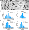
(A) TEM images of nHA and SrHA nanocrystals. Scale bar (200 nm) is the same in the two images, for direct comparison. (B) Length distribution of nHA and SrHA, (C) width distribution of nHA and SrHA.
Figure 3
Figure 3
Alg, Alg0.5nHA, Alg1nHA, Alg0.5SrHA, and Alg1SrHA, at different infill densities. (Top) CAD design of grid with 5% infill density and results obtained for each condition. (Middle) Grids with 25% infill density and results obtained for each condition. Scale bar: 2 mm. (Bottom) Pore appearance obtained for each condition of the 3D bioprinted structures. Scale bar: 250 µm.
Figure 4
Figure 4
Weight variation for Alg, Alg1nHA and Alg1SrHA conditions at different time points.
Figure 5
Figure 5
Calcium and strontium cumulative release from scaffolds containing nHA or SrHA powders as a function of soaking time in HEPES solution. Calcium release is reported from nHA (black ●) and SrHA (red ◼); strontium release from SrHA (blue ▲).
Figure 6
Figure 6
Saos-2 cell viability on pure Alg and Alg1nHA and Alg1SrHA performed by Live/Dead staining and acquired by dual-photon confocal microscopy at 24 h. (A) Volume render; (B) intensity projection (left panel, scale bar 50 µm) and % of cell viability automatically quantified (right panel, medium ± SE, * p < 0.05, n = 3 for Alg and Alg1nHA, and n = 4 for Alg1SrHA).
Figure 7
Figure 7
Metabolically active Saos-2 cells in 3D printed samples at all the considered time points (1, 3, 7, and 14 days) (* p-value ≤ 0.05, ** p ≤ 0.01, *** p ≤ 0.001).
Figure 8
Figure 8
Representative images acquired by optical microscopy of Saos-2 cells embedded in the inks and stained with Live Dead assay at all the considered time points (1, 3, 7, and 14 days). Scale bar 100 µm.
Figure 9
Figure 9
Saos-2 cell viability (quantified by Live/Dead staining, acquisition by optical microscopy) on 3D bioprinted samples composed of pure alginate and alginate + nHA and SrHA particles at different concentrations, over 14 days (* p-value ≤ 0.05).

References

    1. Graziani G., Berni M., Gambardella A., De Carolis M., Maltarello M.C., Boi M., Carnevale G., Bianchi M. Fabrication and characterization of biomimetic hydroxyapatite thin films for bone implants by direct ablation of a biogenic source. Mater. Sci. Eng. C. 2019;99:853–862. doi: 10.1016/j.msec.2019.02.033. - DOI - PubMed
    1. Du Y., Guo J.L., Wang J., Mikos A.G., Zhang S. Hierarchically designed bone scaffolds: From internal cues to external stimuli. Biomaterials. 2019;218:119334. doi: 10.1016/j.biomaterials.2019.119334. - DOI - PMC - PubMed
    1. Bigi A., Boanini E. Functionalized Biomimetic Calcium Phosphates for Bone Tissue Repair. J. Appl. Biomater. Funct. Mater. 2017;15:e313–e325. doi: 10.5301/jabfm.5000367. - DOI - PubMed
    1. Liu Y., Luo D., Wang T. Hierarchical Structures of Bone and Bioinspired Bone Tissue Engineering. Small. 2016;12:4611–4632. doi: 10.1002/smll.201600626. - DOI - PubMed
    1. Fischetti T., Di Pompo G., Baldini N., Avnet S., Graziani G. 3D Printing and Bioprinting to Model Bone Cancer: The Role of Materials and Nanoscale Cues in Directing Cell Behavior. Cancers. 2021;13:4065. doi: 10.3390/cancers13164065. - DOI - PMC - PubMed