. 2023 Jul 24;16(7):1048.
doi: 10.3390/ph16071048.

Renoprotective Effect of Chrysanthemum coronarium L. Extract on Adenine-Induced Chronic Kidney Disease in Mice

Affiliations

Renoprotective Effect of Chrysanthemum coronarium L. Extract on Adenine-Induced Chronic Kidney Disease in Mice

Yi-Seul Kim et al. Pharmaceuticals (Basel). .

Abstract

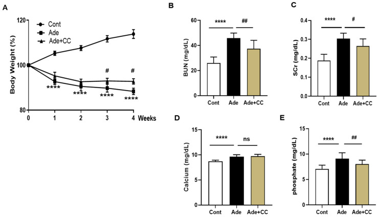
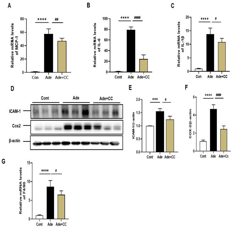
Chronic kidney disease (CKD) gradually leads to loss of renal function and is associated with inflammation and fibrosis. Chrysanthemum coronarium L., a leafy vegetable, possesses various beneficial properties, including anti-oxidative, anti-inflammatory, and antiproliferative effects. In this study, we investigated the renoprotective effect of Chrysanthemum coronarium L. extract (CC) on adenine (AD)-induced CKD in mice. CKD was induced by feeding mice with an AD diet (0.25% w/w) for 4 weeks. Changes in renal function, histopathology, inflammation, and renal interstitial fibrosis were analyzed. The adenine-fed mice were characterized by increased blood urea nitrogen, serum creatinine, and histological changes, including inflammation and fibrosis; however, these changes were significantly restored by treatment with CC. Additionally, CC inhibited the expression of the inflammatory markers, monocyte chemoattractant protein-1, interleukins-6 and -1β, intercellular adhesion molecule-1, and cyclooxygenase 2. Moreover, CC suppressed the expression of the fibrotic markers, type IV collagen, and fibronectin. Furthermore, CC attenuated the expression of profibrotic genes (tumor growth factor-β and α-smooth muscle actin) in AD-induced renal injury mice. Thus, our results suggest that CC has the potential to attenuate AD-induced renal injury and might offer a new option as a renoprotective agent or functional food supplement to manage CKD.

Keywords: Chrysanthemum coronarium L. extract; chronic kidney disease; fibrosis; inflammation.


Conflict of interest statement

The authors declare no conflict of interest.

Figures

Figure 1
Figure 1
Qualitative analysis of bioactive compounds from CC. The sample extract was analyzed using UPLC equipped with an Acquity BEH C18 column (2.1 × 100 mm, 1.7 μm, Waters) at 254 nm (A), and the eluted compounds were ionized by negative electrospray ionization and analyzed using Q-TOF (B). The compounds were identified using their MS spectra (C) and the online database connected to the UNIFI 1.9.2.045 software. Blue symbols are fragment.
Figure 2
Figure 2
CC attenuates adenine (AD)-induced renal injury. Chronic kidney disease (CKD) was induced by AD (0.25% w/w) for 28 days, and CC (100 mg/kg body weight) was administered orally by gavage either alone or with AD. (A) Body weight. (B) BUN level. (C) SCr level. (D) Calcium level. (E) Phosphate level. All data are presented as mean ± standard deviation (SD). n = 8. **** p < 0.0001 vs. Cont group. # p < 0.05, and ## p < 0.01, ns: not significant, vs. Ade group.
Figure 3
Figure 3
CC attenuates AD-induced morphological changes. (A) H&E staining was used for identification and semiquantitative scoring of inflammation. The orange arrows indicate inflammatory cell infiltration. (B) PAS staining was used for identification and semiquantitative scoring of basal membrane atrophy and dilatation. Green arrows indicate examples of atrophic basal membranes and red stars indicate tubule dilation. (C) MT staining was used for identification and semiquantitative fibrosis scoring. The yellow arrows indicate interstitial fibrosis. (D) Histopathology was evaluated from the kidney stained with HE, PAS, and MT. All data are presented as mean ± SD. n = 8. **** p < 0.0001 vs. control group. # p < 0.05, vs. Ade group.
Figure 3
Figure 3
CC attenuates AD-induced morphological changes. (A) H&E staining was used for identification and semiquantitative scoring of inflammation. The orange arrows indicate inflammatory cell infiltration. (B) PAS staining was used for identification and semiquantitative scoring of basal membrane atrophy and dilatation. Green arrows indicate examples of atrophic basal membranes and red stars indicate tubule dilation. (C) MT staining was used for identification and semiquantitative fibrosis scoring. The yellow arrows indicate interstitial fibrosis. (D) Histopathology was evaluated from the kidney stained with HE, PAS, and MT. All data are presented as mean ± SD. n = 8. **** p < 0.0001 vs. control group. # p < 0.05, vs. Ade group.
Figure 4
Figure 4
CC suppresses inflammatory responses in AD-induced renal injury mice. qRT-PCR analysis of MCP-1 (A), IL-6 (B), and IL-1β (C) mRNA expression. Western blotting analysis showing protein expression of ICAM-1 (D,E) and COX-2(D,F). qRT-PCR analysis showing the mRNA expression of F4/80 (G). All data are presented as mean ± SD. n = 8. *** p < 0.001 and **** p < 0.0001 vs. Cont group. # p < 0.05, and ## p < 0.01, #### p < 0.0001, vs. Ade group.
Figure 5
Figure 5
CC reduces extracellular matrix deposition and tubulointerstitial fibrosis in AD-induced renal injury mice. qRT-PCR data showing the mRNA level of type IV collagen (A) and fibronectin (B). All data are presented as mean ± SD. n = 8. ** p < 0.01 and **** p < 0.0001 vs. Cont group. # p < 0.05, and #### p < 0.001, vs. Ade group.
Figure 6
Figure 6
CC reduces the upregulation of TGF-β1 and α-SMA expression in AD-induced renal injury mice. qRT-PCR results of the mRNA levels of TGF-β1 (A) and α-SMA (B). IHC staining showing the expression of α-SMA (C). ImageJ was used for quantification of the α-SMA area (D). All data are expressed as mean ± SD. n = 8. ** p < 0.01, *** p < 0.001, and **** p < 0.0001, vs. Cont group. ### p < 0.001, and #### p < 0.0001 vs. Ade group.

References

    1. Webster A.C., Nagler E.V., Morton R.L., Masson P. Chronic Kidney Disease. Lancet. 2017;389:1238–1252. doi: 10.1016/S0140-6736(16)32064-5. - DOI - PubMed
    1. Sohel B.M., Rumana N., Ohsawa M., Turin T.C., Kelly M.A., Al Mamun M. Renal function trajectory over time and adverse clinical outcomes. Clin. Exp. Nephrol. 2016;20:379–393. doi: 10.1007/s10157-015-1213-0. - DOI - PubMed
    1. Zhang C.-Y., Zhu J.-Y., Ye Y., Zhang M., Zhang L.-J., Wang S.-J., Song Y.-N., Zhang H. Erhuang Formula ameliorates renal damage in adenine–induced chronic renal failure rats via inhibiting inflammatory and fibrotic responses. Biomed. Pharmacother. 2017;95:520–528. doi: 10.1016/j.biopha.2017.08.115. - DOI - PubMed
    1. Sonfack C.S., Nguelefack-Mbuyo E.P., Kojom J.J., Lappa E.L., Peyembouo F.P., Fofié C.K., Nolé T., Nguelefack T.B., Dongmo A.B. The aqueous extract from the stem bark of Garcinia lucida Vesque (Clusiaceae) exhibits cardioprotective and nephroprotective effects in adenine-induced chronic kidney disease in rats. Evid. Based Complement. Alternat. Med. 2021;2021:5581041. doi: 10.1155/2021/5581041. - DOI - PMC - PubMed
    1. Yi H., Huang C., Shi Y., Cao Q., Chen J., Chen X.-M., Pollock C.A. Metformin attenuates renal fibrosis in a mouse model of adenine-induced renal injury through inhibiting TGF-β1 signaling pathways. Fron. Cell Dev. Biol. 2021;9:603802. doi: 10.3389/fcell.2021.603802. - DOI - PMC - PubMed