

Polydopamine-Coated Polycaprolactone Electrospun Nanofiber Membrane Loaded with Thrombin for Wound Hemostasis
- PMID: 37514511
- PMCID: PMC10385294
- DOI: 10.3390/polym15143122
Polydopamine-Coated Polycaprolactone Electrospun Nanofiber Membrane Loaded with Thrombin for Wound Hemostasis
Abstract
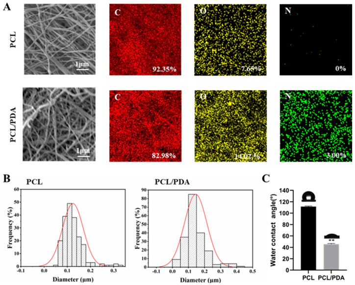
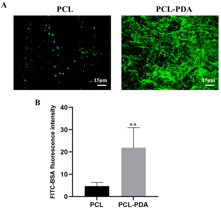
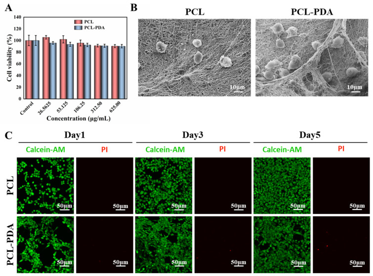
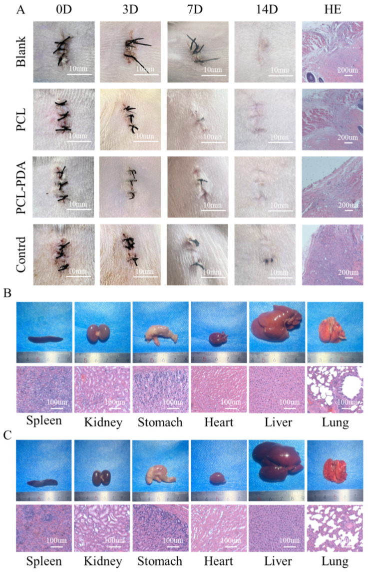
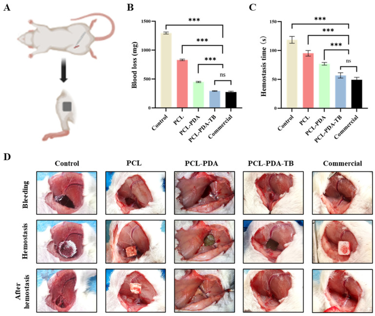
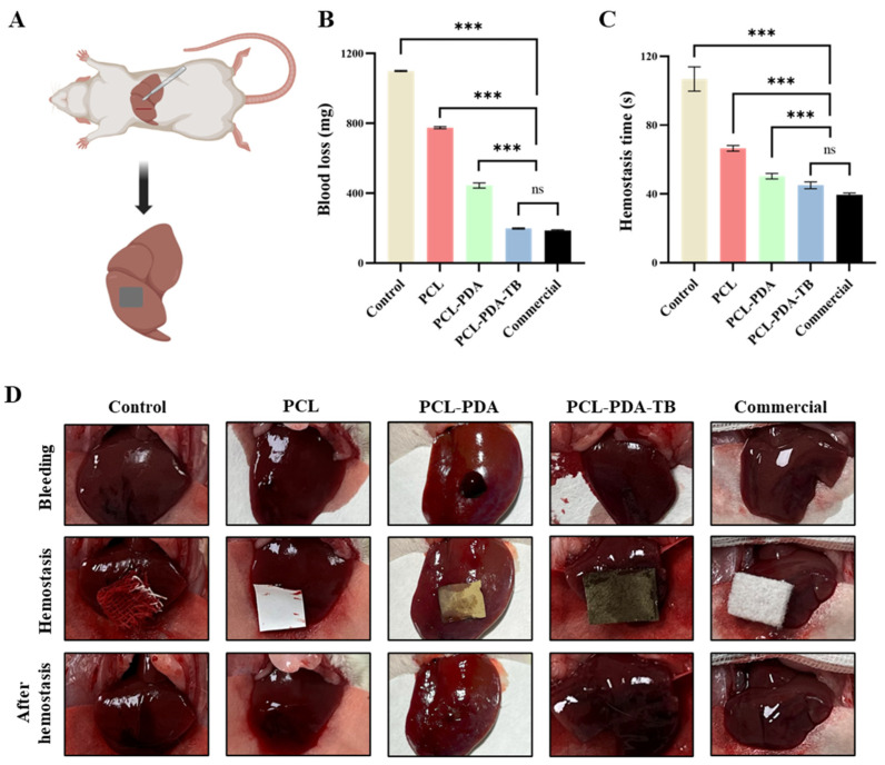
Hemorrhagic shock is the primary cause of death in patients with severe trauma, and the development of rapid and efficient hemostatic methods is of great significance in saving the lives of trauma patients. In this study, a polycaprolactone (PCL) nanofiber membrane was prepared by electrospinning. A PCL-PDA loading system was developed by modifying the surface of polydopamine (PDA), using inspiration from mussel adhesion protein, and the efficient and stable loading of thrombin (TB) was realized to ensure the bioactivity of TB. The new thrombin loading system overcomes the disadvantages of harsh storage conditions, poor strength, and ease of falling off, and it can use thrombin to start a rapid coagulation cascade reaction, which has the characteristics of fast hemostasis, good biocompatibility, high safety, and a wide range of hemostasis. The physicochemical properties and biocompatibility of the PCL-PDA-TB membrane were verified by scanning electron microscopy, the cell proliferation test, the cell adhesion test, and the extract cytotoxicity test. Red blood cell adhesion, platelet adhesion, dynamic coagulation time, and animal models all verified the coagulation effect of the PCL-PDA-TB membrane. Therefore, the PCL-PDA-TB membrane has great potential in wound hemostasis applications, and should be widely used in various traumatic hemostatic scenarios.
Keywords: electrospinning; hemostatic method; polydopamine modification; thrombin; trauma.
Conflict of interest statement
There are no conflict of interest.
Figures

Similar articles
-
Triple PLGA/PCL Scaffold Modification Including Silver Impregnation, Collagen Coating, and Electrospinning Significantly Improve Biocompatibility, Antimicrobial, and Osteogenic Properties for Orofacial Tissue Regeneration.ACS Appl Mater Interfaces. 2019 Oct 16;11(41):37381-37396. doi: 10.1021/acsami.9b07053. Epub 2019 Oct 7. ACS Appl Mater Interfaces. 2019. PMID: 31517483 Free PMC article.
-
Fabrication of thrombin loaded TEMPO-oxidized cellulose nanofiber-gelatin sponges and their hemostatic behavior in rat liver hemorrhage model.J Biomater Sci Polym Ed. 2022 Mar;33(4):499-516. doi: 10.1080/09205063.2021.1992877. Epub 2021 Oct 20. J Biomater Sci Polym Ed. 2022. PMID: 34644247
-
In vitro behavior of tendon stem/progenitor cells on bioactive electrospun nanofiber membranes for tendon-bone tissue engineering applications.Int J Nanomedicine. 2019 Jul 26;14:5831-5848. doi: 10.2147/IJN.S210509. eCollection 2019. Int J Nanomedicine. 2019. PMID: 31534327 Free PMC article.
-
Antimicrobial Activity of 3D-Printed Poly(ε-Caprolactone) (PCL) Composite Scaffolds Presenting Vancomycin-Loaded Polylactic Acid-Glycolic Acid (PLGA) Microspheres.Med Sci Monit. 2018 Sep 30;24:6934-6945. doi: 10.12659/MSM.911770. Med Sci Monit. 2018. PMID: 30269152 Free PMC article.
-
Mussel-inspired nanoparticle composite hydrogels for hemostasis and wound healing.Front Chem. 2023 Mar 30;11:1154788. doi: 10.3389/fchem.2023.1154788. eCollection 2023. Front Chem. 2023. PMID: 37065820 Free PMC article. Review.
Cited by
-
Polydopamine-coated polycaprolactone/carbon nanotube fibrous scaffolds loaded with basic fibroblast growth factor for wound healing.Mater Today Bio. 2024 Aug 6;28:101190. doi: 10.1016/j.mtbio.2024.101190. eCollection 2024 Oct. Mater Today Bio. 2024. PMID: 39221197 Free PMC article.
-
Silk fibroin-gelatine haemostatic sponge loaded with thrombin for wound haemostasis and tissue regeneration.Burns Trauma. 2024 Oct 13;12:tkae026. doi: 10.1093/burnst/tkae026. eCollection 2024. Burns Trauma. 2024. PMID: 39403685 Free PMC article.
-
Electrostimulation combined with biodegradable electroactive oriented nanofiber polycaprolactone/gelatin/carbon nanotube to accelerate wound healing.Mater Today Bio. 2025 Jan 13;31:101490. doi: 10.1016/j.mtbio.2025.101490. eCollection 2025 Apr. Mater Today Bio. 2025. PMID: 39896286 Free PMC article.
-
A sodium alginate - silk fibroin biosponge loaded with thrombin: Effective hemostasis and wound healing.Heliyon. 2024 Mar 15;10(6):e28047. doi: 10.1016/j.heliyon.2024.e28047. eCollection 2024 Mar 30. Heliyon. 2024. PMID: 38524596 Free PMC article.
References
-
- Stewart R.M., Myers J.G., Dent D.L., Ermis P., Gray G.A., Villarreal R., Blow O., Woods B., McFarland M., Garavaglia J., et al. Seven Hundred Fifty-Three Consecutive Deaths in a Level I Trauma Center: The Argument for Injury Prevention. J. Trauma Inj. Infect. Crit. Care. 2003;54:66–71. doi: 10.1097/00005373-200301000-00009. - DOI - PubMed
-
- Muldowney M., Liu Z., Stansbury L.G., Vavilala M.S., Hess J.R. Ultramassive Transfusion for Trauma in the Age of Hemostatic Resuscitation: A Retrospective Single-Center Cohort from a Large US Level-1 Trauma Center, 2011–2021. Obstet. Anesth. Dig. 2023;136:927–933. doi: 10.1213/ANE.0000000000006388. - DOI - PubMed
-
- Sperry J.L., Cotton B.A., Luther J.F., Cannon J.W., Schreiber M.A., Moore E.E., Namias N., Minei J.P., Wisniewski S.R., Guyette F.X. Whole Blood Resuscitation and Association with Survival in Injured Patients with an Elevated Probability of Mortality. J. Am. Coll. Surg. 2023;237:206–219. doi: 10.1097/XCS.0000000000000708. - DOI - PMC - PubMed
Grants and funding
LinkOut - more resources
Full Text Sources
Other Literature Sources
