. 2023 Jul 10;11(7):1224.
doi: 10.3390/vaccines11071224.

Development and Characterization of an In Vitro Cell-Based Assay to Predict Potency of mRNA-LNP-Based Vaccines

Affiliations

Development and Characterization of an In Vitro Cell-Based Assay to Predict Potency of mRNA-LNP-Based Vaccines

Nisarg Patel et al. Vaccines (Basel). .

Erratum in

Abstract

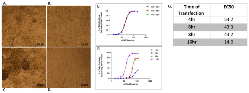
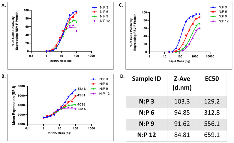
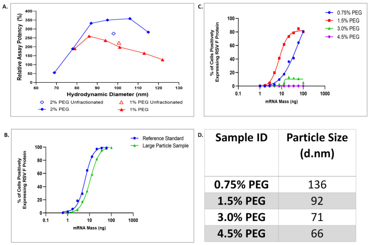
Messenger RNA (mRNA) vaccines have emerged as a flexible platform for vaccine development. The evolution of lipid nanoparticles as effective delivery vehicles for modified mRNA encoding vaccine antigens was demonstrated by the response to the COVID-19 pandemic. The ability to rapidly develop effective SARS-CoV-2 vaccines from the spike protein genome, and to then manufacture multibillions of doses per year was an extraordinary achievement and a vaccine milestone. Further development and application of this platform for additional pathogens is clearly of interest. This comes with the associated need for new analytical tools that can accurately predict the performance of these mRNA vaccine candidates and tie them to an immune response expected in humans. Described here is the development and characterization of an imaging based in vitro assay able to quantitate transgene protein expression efficiency, with utility to measure lipid nanoparticles (LNP)-encapsulated mRNA vaccine potency, efficacy, and stability. Multiple biologically relevant adherent cell lines were screened to identify a suitable cell substrate capable of providing a wide dose-response curve and dynamic range. Biologically relevant assay attributes were examined and optimized, including cell monolayer morphology, antigen expression kinetics, and assay sensitivity to LNP properties, such as polyethylene glycol-lipid (or PEG-lipid) composition, mRNA mass, and LNP size. Collectively, this study presents a strategy to quickly optimize and develop a robust cell-based potency assay for the development of future mRNA-based vaccines.

Keywords: LNP; bioassays; mRNA; potency; potency assays; vaccines.


Conflict of interest statement

Authors Carl Hofmann, Josef Vlasak, John W. Loughney and Malini Mukherjee are employed by Merck Sharp & Dohme LLC., a subsidiary of Merck and Co., Inc., Rahway, NJ, USA and have potential stock ownership in Merck and Co., Inc., Rahway, NJ, USA.

Figures

Figure 1
Figure 1
LNP transfection curves across varying cell lines indicate variable levels of transfection, which improve with the addition of ApoE during transfection. (A) RSV-F protein expression efficiency in Vero, HeLA, HEp-2, A549, CaCo-2 and HepG2 cells was measured by counting percentage of cells positive for RSV-F protein. All cells were transfected for 16 ± 2 h with titration of LNPs starting with 200 ng dose of mRNA in media with 2% FBS. Data were fit using variable slope-four parameter logistics regression (4-PL) model in GraphPad Prism software (version: 6.0). (BG) RSV-F protein expression efficiency following transfection with LNPs in media + 2%FBS, and media + 2%FBS supplemented with ApoE at 4 ug/mL. Hill-slope values [Y = Bottom + (Top − Bottom)/(1 + 10^((LogEC50-X) × Hill-slope)] from 4-PL regression are given for curves obtained with FBS supplemented with ApoE. (B) HeLa cells. (C) Vero cells. (D) Hep-2 cells. (E) CaCo-2 cells. (F) A549 cells. (G) HepG2 cells. (H) Representative immunofluorescence images of HepG2, Vero and A549 cells without ApoE and (I) with ApoE shown at 72 (top row) and 200 ng/mL (bottom row) of mRNA dose representing the bottom and top of the dose response curve, respectively.
Figure 2
Figure 2
Optimization of cell plating parameters and transfection kinetics. Morphological characteristics of HepG2 monolayers on tissue culture plates and collagen I-coated plates at different cell seeding densities along with transfection kinetics of RSV-F protein encoding mRNA encapsulated within LNP. (A) HepG2 cells seeded at 7.5 × 10⁴ cells/well on tissue culture plates. (B) HepG2 cells seeded at 7.5 × 10⁴ cells/well on collagen I plates. (C) HepG2 cells seeded at 5.0 × 10⁴ cells/well on collagen I plates. (D) HepG2 cells seeded at 2.5 × 10⁴ cells/well on collagen I plates. (E) Comparison of RSV-F protein expression efficiency following transfection with LNPs at seeding densities of 2.5, 3.0 and 3.5 × 10⁴ cells/well. (F) RSV-F expression efficiency at 4, 6, 8 and 16 h post-transfection and (G) EC50 of dose–response is tabulated in ng of mRNA at each transfection time period.
Figure 3
Figure 3
Potency assay is stability-indicating with the ability to discriminate changes in mRNA content in relation to LNP. (A) RSV-F expression efficiency for LNPs with N/P ratios of 3, 6, 9 and 12 with transfection and protein expression graphed as a function of mRNA mass. (B) RSV-F mean fluorescence per cell for LNPs with N/P ratios of 3, 6, 9, and 12 graphed as a function of mRNA mass. (C) RSV-F expression efficiency for LNPs with N/P ratios of 3, 6, 9, and 12 graphed as a function of lipid mass, to approximate particle number. (D) LNP particle size for differing N/P ratios and associated EC50 values for Figure 3C.
Figure 4
Figure 4
LNP transfection in HepG2 cells is optimal within a narrow size and PEGylation range. (A) Percent relative assay potency for 1% and 2% PEG-DMG unfractionated samples compared to samples that underwent density gradient centrifugation resulting in 6 differing size fractions. (B) RSV-F expression efficiency for sample lots with differing mean LNP size. (C) RSV-F expression efficiency for LNPs with PEG composition of 0.75, 1.5, 3.0 and 4.5% and (D) associated particle size.

References

    1. Zhang C., Maruggi G., Shan H., Li J. Advances in mRNA Vaccines for Infectious Diseases. Front. Immunol. 2019;10:594. doi: 10.3389/fimmu.2019.00594. - DOI - PMC - PubMed
    1. Jackson N.A.C., Kester K.E., Casimiro D., Gurunathan S., DeRosa F. The promise of mRNA vaccines: A biotech and industrial perspective. npj Vaccines. 2020;5:11. doi: 10.1038/s41541-020-0159-8. - DOI - PMC - PubMed
    1. Wu F., Zhao S., Yu B., Chen Y.M., Wang W., Song Z.G., Hu Y., Tao Z.W., Tian J.H., Pei Y.Y., et al. A new coronavirus associated with human respiratory disease in China. Nature. 2020;579:265–269. doi: 10.1038/s41586-020-2008-3. - DOI - PMC - PubMed
    1. Pardi N., Hogan M.J., Porter F.W., Weissman D. mRNA vaccines—A new era in vaccinology. Nat. Rev. Drug Discov. 2018;17:261–279. doi: 10.1038/nrd.2017.243. - DOI - PMC - PubMed
    1. Tam Y.Y., Chen S., Cullis P.R. Advances in Lipid Nanoparticles for siRNA Delivery. Pharmaceutics. 2013;5:498–507. doi: 10.3390/pharmaceutics5030498. - DOI - PMC - PubMed

LinkOut - more resources