. 2023 Jun 23;15(7):1424.
doi: 10.3390/v15071424.

Structure of Bovine CD46 Ectodomain

Affiliations

Structure of Bovine CD46 Ectodomain

Hazel Aitkenhead et al. Viruses. .

Abstract

CD46, or membrane cofactor protein, is a type-one transmembrane protein from the complement regulatory protein family. Alongside its role in complement activation, CD46 is involved in many other processes, from T-cell activation to reproduction. It is also referred to as a pathogen magnet, because it is used as a receptor by multiple bacteria and viruses. Bovine CD46 (bovCD46) in particular is involved in bovine viral diarrhoea virus entry, an economically important disease in cattle industries. This study presents the X-ray crystallographic structure of the extracellular region of bovCD46, revealing a four-short-consensus-repeat (SCR) structure similar to that in human CD46. SCR1-3 are arranged linearly, while SCR 4 has a reduced interface angle, resulting in a hockey stick-like appearance. The structure also reveals the bovine viral diarrhoea virus interaction site in SCR1, which is likely to confer pestivirus specificity for their target host, CD46. Insights gained from the structural information on pestivirus receptors, such as CD46, could offer valuable guidance for future control strategies.

Keywords: BVDV; CD46; X-ray structure; bovine viral diarrhoea virus; pestivirus; type I membrane protein; virus receptor.


Conflict of interest statement

The authors declare no conflict of interest.

Figures
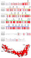
Figure 1
Figure 1
Amino acid conservation between CD46 proteins. Top—Sequence alignment of bovCD46 with CD46 from humans, pigs, and sheep generated using Clustal Omega [18] and ESPript web service [19]. Conserved residues are highlighted in red and similar residues are in red text. Secondary structure from bovCD46 is shown above and SCR beta sheet labelling has been added. The sequence numbering for each SCR has been highlighted as: SCR1—orange, SCR2—yellow, SCR3—green, and SCR4—blue. Green numbering below shows cysteines involved in disulphide bonds and glycosylated asparagines are marked with a blue triangle. GenBank accession numbers: Bovine— Q6VE48, Human—P15529, Porcine—O02839, and Ovine—W5PWS0. Bottom—Structure of human CD46_SCR1-4, with the surface representing sequence conservation on a scale of non-conserved white residues to conserved red residues.
Figure 2
Figure 2
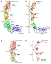
Overall structure of the bovCD46 extracellular domain. (A) Structure of SCR1-4 monomer in a drawing with the surface shown. SCR1 at the N-terminus is orange, SCR2 is yellow, SCR3 is green, and SCR4 at the C-terminus is blue. Cysteines and, therefore, disulphide bridges are shown in magenta balls and sticks. Domain boundaries are marked with the N-terminal boundary at the top left of the domain and the C-terminal at the bottom right. (B) Drawing of SCR1-4 monomer without surface with position in relation to marked membrane and labelled SCRs. The glycan residues are shown in red sticks and the asparagine residues, which are glycosylated in at least one monomer, are labelled and shown in black sticks. (C) As in (A), but for the SCR1-2 monomer. (D) As in (B), but for the SCR1-2 monomer.
Figure 3
Figure 3
Structural comparison of individual SCR domains, SCR domain from bovCD46_SCR1-4 shown as a drawing with cysteines coloured magenta. (A) Drawing of SCR1 of bovCD46_SCR1-4 coloured in orange with the BVDV interaction site highlighted in cyan. (B) Drawing of SCR1 of bovCD46_SCR1-4 coloured in orange superposed with SCR2 in yellow, RMSD 4.1 Å over 60 residues. (C) Drawing of SCR1 of bovCD46_SCR1-4 coloured in orange superposed with SCR3 in green, RMSD 4.6 Å over 64 residues. (D) Drawing of SCR1 of bovCD46_SCR1-4 coloured in orange superposed with SCR4 in blue, RMSD 3.7 Å over 60 residues.
Figure 4
Figure 4
Structural comparison of bovCD46 and human CD46. (A) Drawing of bovCD46_SCR1-4 coloured purple with the BVDV interaction site highlighted in orange, a drawing of the human CD46 is superposed in blue (PDB:3O8E). (B) Closeup view of residues (shown as sticks) involved in the BVDV interaction site—the colour scheme is identical to (A). (C) Surface representation of bovCD46_SCR1-4 coloured by sequence conservation on a scale of non-conserved white residues to conserved red residues. BVDV interaction site is outlined in black and below is an excerpt from CD46 alignment focusing on BVDV interaction site peptides, both generated using ESPript web service [19].
Figure 5
Figure 5
Crystallographic dimer found in bovCD46. (A) Model of bovCD46_SCR1-2 with both monomers from the asymmetric unit shown with a surface representation in blue and yellow. (B) Model of bovCD46_SCR1-4 with both monomers from the asymmetric unit shown with a surface representation in purple and green (the green monomer is a copy of the purple monomer superposed in the correct position, as SCR4 was not resolved in this chain). (C) Hydrogen bonding patch at the dimer interface of bovCD46_SCR1-2 with residues in hydrogen bonding distance shown in cyan sticks and the potential hydrogen bonds also in cyan. (D) Dimer interface of bovCD46_SCR1-4 with residues in hydrogen bonding distance shown in cyan sticks and the potential hydrogen bonds also in cyan.

References

    1. Seya T., Turner J.R., Atkinson J.P. Purification and characterization of a membrane protein (gp45-70) that is a cofactor for cleavage of C3b and C4b. J. Exp. Med. 1986;163:837–855. doi: 10.1084/jem.163.4.837. - DOI - PMC - PubMed
    1. Liszewski M.K., Post T.W., Atkinson J.P. Membrane Cofactor Protein (MCP or CD46): Newest Member of the Regulators of Complement Activation Gene Cluster. Annu. Rev. Immunol. 1991;9:431–455. doi: 10.1146/annurev.iy.09.040191.002243. - DOI - PubMed
    1. Persson B.D., Schmitz N.B., Santiago C., Zocher G., Larvie M., Scheu U., Casasnovas J.M., Stehle T. Structure of the Extracellular Portion of CD46 Provides Insights into Its Interactions with Complement Proteins and Pathogens. PLoS Pathog. 2010;6:e1001122. doi: 10.1371/journal.ppat.1001122. - DOI - PMC - PubMed
    1. Arlaud G.J., Barlow P.N., Gaboriaud C., Gros P., Narayana S.V.L. Deciphering complement mechanisms: The contributions of structural biology. Mol. Immunol. 2007;44:3809–3822. doi: 10.1016/j.molimm.2007.06.147. - DOI - PubMed
    1. Post T.W., Liszewski M.K., Adams E.M., Tedja I., Miller E.A., Atkinson J.P. Membrane cofactor protein of the complement system: Alternative splicing of serine/threonine/proline-rich exons and cytoplasmic tails produces multiple isoforms that correlate with protein phenotype. J. Exp. Med. 1991;174:93–102. doi: 10.1084/jem.174.1.93. - DOI - PMC - PubMed

Publication types

Substances

LinkOut - more resources